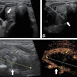
(Ultrasound Review) According to researchers at the Vanderbilt University Medical Center in Nashville, color Doppler ultrasound may be useful in differentiating benign polyps and endometrial carcinoma from hematrometra in women with enlarged endometria.
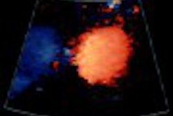
"Color Doppler sonography currently provides a means to distinguish thickened endometrial interfaces due to endometrial polyps or carcinoma from hematometra, in which flow would not be shown, thereby distinguishing those patients who need simple drainage of the luminal contents from those requiring dilation and curettage or hysteroscopy," they reported in retrospective study in the August issue of Journal of Ultrasound in Medicine.
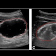
The ultrasound findings of 15 women with abnormal bleeding were compared with the histologic findings of endometrial masses. Women with a bilayered endometrial thickness greater than 6mm and abnormal bleeding were included in the study. Ten women were postmenopausal and five were premenopausal. Using transvaginal transducers ranging in frequency from 8-4MHz they determined the number of main and secondary blood vessel using both color and power Doppler flow. A determination of the microvessel density from the surgical specimen also was made.
They reported that "of the 15 women studied, 10 had benign polyps, four had carcinoma, and one had a lipoleiomyoma." Color Doppler showed vascularity in both benign and malignant endometrial masses, and the presence of vessels on color flow did not relate to the malignancy risk. Although the number of vessels shown on color Doppler correlated with malignancy, it did not correlate with microvessel density.
"This lack of correlation may be due to subjective selection of areas to count microvessels rather than the ability of color Doppler to detect smaller vessels," they suggested.
Results indicated that although color Doppler was useful for further assessment of a thickened endometrium, this limited study suggests it may not be sufficiently accurate to differentiate benign from malignant lesions. The authors suggested that a similar study using a larger patient population would be beneficial in establishing the role of color Doppler in diagnosing endometrial pathology, particularly in women on hormone replacement therapy or Tamoxifen.
Color Doppler sonography of endometrial massesFleischer, A. C. et al
Department of radiology and radiological sciences, Vanderbilt University Medical Center, Nashville, TN
J Ultrasound Med 2002 August; 21:861-865
By Ultrasound Review
September 18, 2002
Copyright © 2002 AuntMinnie.com
