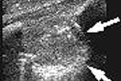
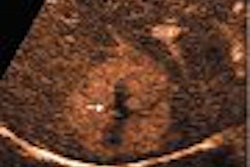
Biomedical engineers at Duke University's Pratt School of Engineering have created a new 3D ultrasound cardiac imaging probe.
The 3D transesophageal echocardiography (TEE) probe uses the outer casing of a commercially available 2D TEE probe to house the new technology, as the casing design already has been tested and approved for use, according to the Durham, NC-based institution.
In addition to its potential applications for cardiac imaging, the new probe can also be used to image the esophagus, rectum, colon, and prostate, the university said.
The probe generates ultrasound at 5 million vibrations per second, and features 504 sensors. In animal testing, the unit has generated high-contrast images of the whole heart, and positioned catheters and ablation devices at the same time, Duke engineers said.
By AuntMinnie.com staff writers
May 27, 2005
Related Reading
Duke group delivers diagnostic CT and PET on PET/CT, March 24, 2005
Singapore in medical school alliance with Duke, February 1, 2005
Cardiac MR center finds success with 'one-stop shopping' model, January 25, 2005
New method provides individualized cancer prognosis, April 28, 2004
Copyright © 2005 AuntMinnie.com


