Automated breast ultrasound (ABUS) has proved to be as effective as handheld ultrasound (HHUS) for identifying lesions, according to a pilot study out of the Elizabeth Wende Breast Clinic in Rochester, NY. The technique has potential as a screening adjunct to mammography, according to the lead author.
"The pilot study was performed on diagnostic patients to examine its ability to visualize known areas of interest and its equivalence to handheld ultrasound," explained Dr. Stamatia Destounis in an e-mail to AuntMinnie.com. "Handheld ultrasound, performed by the radiologist, will continue to be the diagnostic tool."
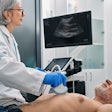
Destounis, a clinical associate professor at the University of Rochester, presented the results of her group's research at the 2005 RSNA in Chicago. For this study, images of known regions of interest were collected by five radiologists using traditional ultrasound. Both benign and malignant lesions were included, and were indicated with a skin marker during HHUS. Hard-copy mammograms and HHUS images were compared with soft-copy ABUS images.
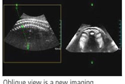
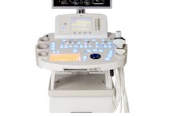
For ABUS, the images were acquired in three views with the patient in a supine position. The ABUS system (SomoVu, U-Systems, San Jose, CA) had a scanning frequency of 10 MHz and a 14.5-cm field-of-view. The scanning depth was 5 cm.
"The unit has automated settings/scanning parameters, which are useful," Destounis said, explaining an advantage of ABUS over operator-dependent HHUS. Also, "ABUS can be performed by a trained technologist (rather than a radiologist). The ABUS can provide additional information to the radiologist.... This can save time as the radiologist will interpret the ABUS scan, but not perform it themselves."
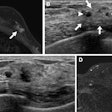
According to the results, in 40 patients ABUS identified 41 lesions, including cysts, fibroadenomas, and lesions that were highly suspicious for cancer. All the cancers (15/15) were found with ABUS on at least one view. Core biopsy proved 13 of the 15. The mean size of these lesions was 14 mm and 54% were nonpalpable.
Many of these patients (93%) had heterogeneously dense breasts, which makes ABUS particularly useful in women with dense breasts, Destounis said.
The concordance between mammography and HHUS versus mammography and ABUS came in at 91%. Based on ABUS results, two cases were downgraded from BI-RADS 3 to 2, while one case was upgraded from BI-RADS 3 to 4. However, none of these cases were cancer.
Destounis said the next phase of research will be a larger clinical trial to test ABUS' performance as a screening tool in conjunction with mammography.
The exact role of ultrasound in breast cancer screening is still under review by the radiology community. As of December 2005, the American College of Radiology Imaging Network (ACRIN 6666) protocol was closer to achieving its patient enrollment goal (2,706 of 2,808). This multicenter trial will assess whole-breast ultrasound for screening in a high-risk population.
In a recent study, Dr. Christiane Kuhl and colleagues out of Bonn, Germany, stated that the combination of ultrasound with mammography seemed insufficient for early diagnosis of breast cancer in high-risk women (Journal of Clinical Oncology, November 20, 2005, Vol. 23:33, pp. 8469-8476).
Previously, Dr. Wendie Berg, Ph.D., and co-investigators found that diagnostic ultrasound was more sensitive in mammography in dense breasts, although there was the risk of overestimating tumor extent with ultrasound (Radiology, December 2004, Vol. 233:3, pp. 830-849).
Still, the promise of ABUS may lie in digging up previously unseen cancers. "We see it as an additional tool in patients (in which) mammography's performance is lacking (in dense-breasted women)," Destounis said.
By Shalmali Pal
AuntMinnie.com staff writer
January 13, 2006
Related Reading
Lesion size, diameter, and location affect breast US interpretation, November 28, 2005
3D imaging creates paradox for ultrasound vendors, November 9, 2005
US-guided optical breast imaging separates benign from malignant tumors, October 13, 2005
Copyright © 2006 AuntMinnie.com


