Tuesday, November 27 | 3:40 p.m.-3:50 p.m. | SSJ01-05 | Arie Crown Theater
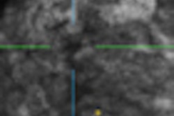
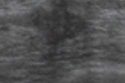
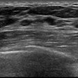
An echogenic "white wall" in a hypoechoic lesion visualized on automated breast ultrasound (ABUS) suggests a benign lesion, and patients may not need to be recalled for further evaluation, according to researchers from George Washington University Medical Center.Dr. Rachel Brem and colleagues observed in their practice that benign cystic lesions have a characteristic white, echogenic wall. They sought to determine how often benign cystic lesions have this characteristic, and compare the frequency with that of cancers when imaged with ABUS.
Their study included 58 women with 64 biopsy-proven cysts and 44 women with 48 biopsy-proven cancers, all of which were imaged with automated breast ultrasound. Of the 64 cysts, 39 (61%) demonstrated the white wall characteristic, while none of the 48 cancers did, Brem's group found.
Based on the results, the team concluded that this characteristic can differentiate benign lesions from malignant ones and reduce recalls of patients with hypoechoic lesions that don't conform to simple cyst criteria.

