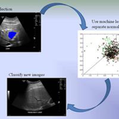
Researchers have created an algorithm for computer-aided detection (CAD) of ultrasound scans that can diagnose several diseases at once using quantitative texture analysis, according to an article in the Journal of Medical Imaging.
Investigators used CAD to analyze regions of interest on ultrasound images for three conditions: craniosynostosis, steatosis, and adenomyosis. The algorithm achieved 72% accuracy for steatosis, 77% accuracy for adenomyosis, and 89% accuracy for craniosynostosis.
The accuracy of the CAD framework is comparable to that of expert readers for steatosis and far exceeds current methods for adenomyosis and craniosynostosis, wrote lead author Jie Ying Wu and colleagues from Brown University (J Med Imaging, January 25, 2016, Vol. 3:1).
 Jie Ying Wu from Brown University.
"We have created a simple and adaptable framework for computer-aided diagnosis that has been shown to be effective in various clinical applications," Wu told AuntMinnie.com by email. "The results show that machine-learning techniques can be used to improve patient care now. With almost no additional cost to the current clinical pipeline, we can have an algorithm that will act as a second opinion and flag cases that are suspicious for a disease."
A challenge to CAD?
The use of ultrasound to detect craniosynostosis, steatosis, and adenomyosis is no mystery, owing to the modality's speed, safety, and low cost. However, operator dependence, interreader variability, and machine settings can interfere. CAD can improve confidence in ultrasound diagnosis through quantitative analysis, the authors wrote.
Their study sought to provide a "straightforward framework for developing a library of tools specific to various image assessment tasks that can increase confidence in diagnosis, and increase detection rates to provide earlier intervention," they wrote.
Steatosis. Steatosis, or abnormal lipid retention, is a promising target for CAD because it occurs in almost a third of the adult population, and although it's reversible with early treatment, long-term disease can lead to severe liver conditions. Ultrasound is adequate for detection, but differences between diseased and healthy tissue can be subtle, leading to frequent biopsies.
Texture changes in the liver due to steatosis have revealed that liver tissues turn from smooth and dark to coarse and grainy as fat content increases. But the changes aren't clear enough to diagnose confidently with ultrasound alone, and creating a dependable framework to systematically quantify ultrasound images is still an open problem, Wu and colleagues wrote.
Adenomyosis. Adenomyosis, defined as the presence of ectopic endometrial tissue within the myometrium, occurs in anywhere from 5% to 70% of women. MRI is the gold standard for detection, but ultrasound is often used because it's cheaper and more available when women present with symptoms such as heavy menstrual bleeding, cramps, and cyclical pain.
A previous study showed that textural features such as subendometrial linear striation and heterogeneous myometrium are indicative of the disease. Wu and colleagues applied the same detection framework as for steatosis to the CAD algorithm.
Craniosynostosis. Craniosynostosis is a premature fusing of sutures in an infant's head that occurs in 1 in 2,000 births; the resulting elevated cranial pressure can affect brain development, the authors wrote. The prognosis is good if an intervention is performed early, ideally between 6 weeks and 10 months of age.
Study methods
To test the CAD algorithm for each condition, the researchers applied it to three separate groups of patients. Manual measurements were acquired on the same ultrasound scanner at every 10° for each image; shape features were used for parameterization.
Wu and colleagues applied CAD to scans of 288 patients with steatosis and 88 patients with adenomyosis. For these two groups, the diagnosis was confirmed with MRI and biopsy. The craniosynostosis dataset consisted of 22 CT-confirmed cases and 22 controls.
For adenomyosis, the radiologist was not blinded and chose regions that were indicative of adenomyosis or normal tissue based on MRI guidelines for texture analysis. For craniosynostosis, the team chose standardized cross-sectional axial cranial images at the plane used to measure biparietal diameter.
Machine-learning classifiers and decision trees were used in all techniques to classify parameters as "sick or not sick," with results based on majority classification. Subclassifiers called trees were used to classify each region as normal or abnormal based on chosen parameters, according to the authors.
| CAD results by clinical condition | |||
| Target | Accuracy | Area under the receiver operator characteristics (ROC) curve | p-value |
| Steatosis | 72.74% | 0.71 | < 0.0001 |
| Adenomyosis | 77.27% | 0.77 | < 0.0001 |
| Craniosynostosis | 88.63% | 0.89 | < 0.0006 |
CAD's accuracy equals that of expert readers for steatosis and exceeds standard clinical diagnostic accuracy for adenomyosis and craniosynostosis, the group wrote. What's more, the CAD algorithm differs from previous techniques in that it uses a machine-learning design that can be trained by analyzing datasets of clinical images and tuned to provide quantitative clinical decision support.
"I believe that these tools can help doctors by flagging high-risk cases," Wu told AuntMinnie.com. "Doctors have a vast amount of additional knowledge like patient history that our algorithm cannot use. If the algorithm can flag some of the more obvious cases from images alone, it could allow doctors to focus on the more difficult cases and help more patients."
 Image courtesy of Jie Ying Wu.
Diagnosing steatosis earlier could prevent more severe symptoms, while adenomyosis detection could help more patients get treated, the authors wrote. Identifying at-risk cases of craniosynostosis earlier could help with treatment planning and counseling.
Interobserver variability
The current study is larger and the dataset more heterogeneous than similar efforts in any of the targeted conditions, according to the authors.
They are considering the addition of image normalization techniques, but this may be less promising than initially thought as simple normalizations don't offer much improvement.
"It is exactly the nonnormal features that we want to identify here, so it's hard not to throw out the baby with the bathwater," said senior author Derek Merck, PhD.
Also on the table is the use of CAD to analyze shear-wave elastography exams, which measure tissue stiffness. Current methods present unique challenges in ultrasound, Wu said.
"Two operators scanning the same patient can produce drastically different-looking images," she said. "Shear-wave elastography should provide a more consistent and generalizable measurement across patients. Also, we are interested in extracting the raw ultrasound measurements to see if our method can learn better from the information that would otherwise be lost in postprocessing."


