Ultrasound may be able to identify women who are at risk of giving birth prematurely, according to a team of research nurses at the University of Illinois at Chicago (UIC).
More than 440,000 babies are born prematurely in the U.S. each year -- that is, before 37 weeks of pregnancy, according to UIC professor of nursing Barbara McFarlin, PhD, and colleagues. Premature births can result in behavioral and neurological disorders and physical problems such as pneumonia and meningitis, in addition to increasing healthcare costs due to longer hospital stays.
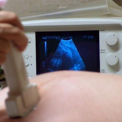
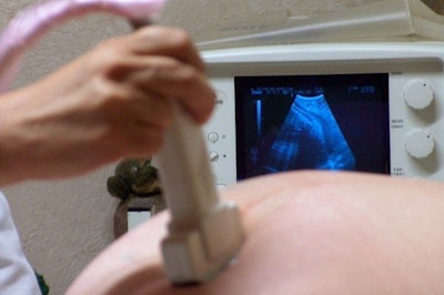
 A UIC researcher using imaging to identify women at risk of giving birth prematurely. Image courtesy of UIC.
Although there is currently no way to predict premature birth, some clinicians associate a shortened cervix with increased risk. In previous research in pregnant rats and women, McFarlin and colleagues used ultrasound to detect collagen tissue changes in the cervix, finding that collagen changes appeared in the cervix before it shortened.
"At 17 to 20 weeks of pregnancy we were able to predict who was going to deliver preterm," McFarlin said in a statement released by UIC. "We found that before the length of the cervix shortens, the microscopic tissue structure has to change and the collagen remodels."
The researchers are also conducting a new study with the support of a five-year, $2.84 million grant from the U.S. National Institute of Child Health and Human Development. McFarlin's team will use ultrasound to detect collagen tissue changes in the cervix in 800 pregnant women divided into three groups:
- Women who previously had a premature baby
- Women who at 20 weeks have a shortened cervix
- A control group at low risk for premature birth
Study participants will undergo an ultrasound examination of the cervix twice: once at 20 weeks of pregnancy and again at 24 weeks.
"By recognizing which women are at risk, healthcare professionals could provide early interventions [and] treatments and closely monitor these treatments to prevent preterm birth or to improve health outcomes," McFarlin said.


