Purdue University researchers are developing a biomedical imaging system that combines optical and ultrasound technology to help clinicians better diagnose disease, according to research published August 28 in Photoacoustics.
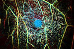
Called photoacoustic tomography, the technology converts optical energy into an acoustic signal by sending pulsed light into body tissue, causing the tissue to expand and creating an acoustic response that can be detected on ultrasound. It produces real-time compositional information of body tissue without the use of contrast agents and with better depth penetration than conventional optical techniques, the university said.
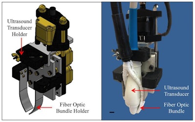
The system consists of a motorized photoacoustic holder that allows users to maneuver fiber-optic bundles and improve the light penetration depth and signal-to-noise ratio of the images. It can be used to detect cardiovascular disease, diabetes, and cancer, as well as to assess lipid deposition within arterial walls, measure cardiac tissue damage, and help surgeons ensure they've removed all cancer from a patient after tumor biopsy, according to Craig Goergen, PhD, from Purdue's Weldon School of Biomedical Engineering.
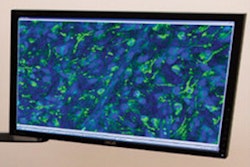
 Purdue University researchers are developing a biomedical imaging system that combines optical and ultrasound technology to improve the diagnosis of life-threatening diseases. Image courtesy of Purdue Research Foundation.
"[Diagnosing disease] at an earlier time can lead to improved patient care," Goergen said a statement released by the university. "We are in the process now of trying to use this enhanced imaging approach [for] a variety of different applications."


