Wednesday, December 4 | 12:45 p.m.-1:15 p.m. | BR278-SD-WEB5 | Lakeside, BR Community, Station 5
In this poster presentation, researchers will discuss how ultrasound tomography is a feasible and accurate tool for characterizing stiffness of breast lesions.The findings offer an alternative to handheld ultrasound, which does provide tissue stiffness and elasticity information but can be operator dependent. In addition, automated breast ultrasound (ABUS) does not measure elasticity -- which makes automated ultrasound tomography an attractive option, noted presenter Neb Duric, PhD, the chief technology officer of Delphinus Medical Technologies, and colleagues.
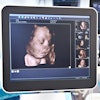
Ultrasound tomography produces 3D sound maps that identify tissue types and measure lesion volumes. Duric's team evaluated imaging of tissue stiffness throughout the breast using the technology in 50 women who had undergone handheld ultrasound; the group used pathology and radiology reports to verify the ultrasound tomography results.
Of the 50 women, 16 had cancers, 16 had fibroadenomas, and 18 had cysts. Ultrasound tomography was able to measure tissue stiffness throughout the breast and characterize lesion stiffness in all 50 patients, with better performance compared with handheld ultrasound, the researchers found: 15 of the 16 cancers were characterized as stiff using the ultrasound tomography method, compared with eight using the handheld technique.
Ultrasound tomography has the potential to improve patient care, according to Duric's group.
"This approach measures tissue stiffness throughout the volume of the breast, something that is currently not possible with other ultrasound devices," the team concluded. "The addition of this information in a screening environment has the potential to reduce call backs and biopsies by utilizing stiffness to improve specificity."


