Ultrasound can more accurately map arrhythmic sites in the heart for improved success of ablation procedures than the more commonly used electrocardiogram (ECG), researchers found.
Typically, catheter ablation is used to kill off misfiring regions, and ECG is often used to identify those regions. However, ECG mapping accuracy is limited and results in inaccurate or incomplete ablation, according to Elisa Konofagou, PhD, of Columbia University.
To solve that problem, Konofagou and her team developed an ultrasound-based technique, electromechanical wave imaging (EWI), to better target those misfiring regions.
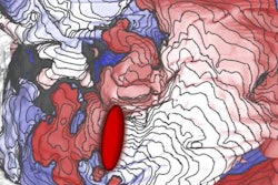
Using ultrasound, EWI creates 3D cardiac maps that noninvasively pinpoint the electromechanical activity causing arrhythmias EWI can also be used during procedures to guide ablation catheters.
Konofagou and colleagues put EWI to the test in a study published in Science Translational Medicine (March 25, 2020). They compared EWI with standard 12-lead ECG on 51 patients undergoing catheter ablation.
They found EWI correctly predicted 96% of arrhythmia locations compared with 71% for ECG. Konofagou and colleagues are planning a larger, long-term clinical trial to further evaluate the technique.


