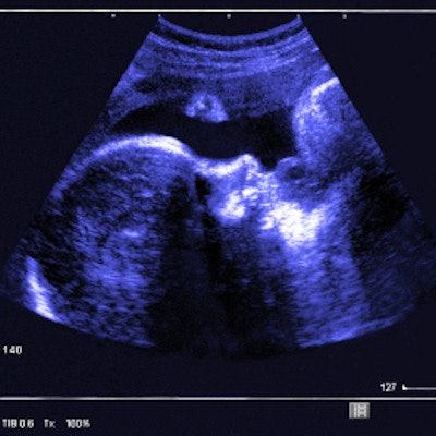
Ultrasound can "moderately" help predict if operative vaginal delivery will be difficult by estimating the head position and station of the fetus, according to research published February 19 in the Journal of Gynecology Obstetrics and Human Reproduction.
A team led by Dr. Alix Plurien from Chu De Lille in France wrote that ultrasound can find factors linked to higher risk of difficult delivery, which could call for an experienced obstetrician to help with delivery procedures that come with risk.
"Nowadays, ultrasound assessment before operational vaginal delivery represents an important tool in a delivery room," Plurien and colleagues wrote. "For the past 15 years, the use of ultrasound has been proposed for predicting the mode of delivery and complications during delivery."
It's common to use digital examination to estimate the position and station of the fetal head during labor. But previous research suggests that digital examination is subjective and has an error rate ranging from 20% to 70%. This can result in complications during delivery, such as abnormal fetal heart rate or failure to progress.
"This error risk is higher after one or more hours of active pushing," the study authors wrote.
Estimating the fetal head station in the pelvis is a must when operational vaginal delivery is needed. The researchers have touted ultrasound's ability to measure the degree of fetal head engagement to make up for the faults of digital examination.
Plurien and colleagues wanted to find out whether ultrasound could accurately assess fetal head position measurement, as well as the distance between the fetal head and perineum. They wanted to see if this could predict a difficult delivery more than conventional digital examination.
The researchers looked at data from 286 operational vaginal deliveries, 65 of which were deemed difficult. Delivery time was longer in the difficult delivery group at 13.8 minutes on average than in the control group at seven minutes (p < 0.001).
| Fetal factors linked to difficult operational vaginal delivery | ||
| Factors | Odds ratio | p-value |
| Posterior and transverse positions | 2.931 | p = 0.0003 |
| Head-perineum distance with soft tissue compression (threshold of 17 mm) | 2.594 | p = 0.0114 |
| Head-perineum distance without soft tissue compression (threshold of 37 mm) | 2.327 | p = 0.0080 |
The team found that the area under the curve (AUC) for predicting difficult delivery according to fetal position from ultrasound was 0.66. Prediction from digital examination, meanwhile, was 0.62. The researchers noted that there was no significant difference between the two methods, which also showed an agreement of 66.8% when measuring fetal head position.
The study authors also found that the AUC of the head-to-perineum distance without soft-tissue compression was 0.59 when measuring fetal station. Without compression, the AUC was 0.60. The team wrote that this means the data do not guide clinicians on whether to perform head-perineum distance measurements with or without pressure.
The group highlighted their evaluation of ultrasound in real practice as one of the study's strengths. Ultrasound before delivery was not used in 16.4% of cases due to conditions such as head on perineum, fetal bradycardia, and unavailability of the ultrasound scanner.
The researchers called for future studies focusing on handling ultrasound and better defining its indications and thresholds in specific populations.
"We did not evaluate intra- and interrater variability for head-perineum distance measurements, which would be interesting to compare in future studies, especially in obese patients," the authors wrote.


