Combining automated breast ultrasound (ABUS) with contrast-enhanced ultrasound (CEUS) could be useful in predicting breast cancer treatment response, according to research published April 24 in Ultrasound in Medicine & Biology.
A team led by Yongwei Xie from Harbin Medical University Cancer Hospital in China found that the combined method yielded better diagnostic accuracy than each individual modality for predicting success of neoadjuvant chemotherapy.
"This model could be used clinically to optimize the treatment of patients with breast cancer," Xie and colleagues wrote.
Ultrasound may be a safe alternative to breast MRI in assessing response to neoadjuvant chemotherapy and guiding treatment interventions. MRI can be hard to access in certain geographic areas and has longer scanning times and higher costs. However, recent technological advances in ultrasound could enable the modality to go toe-to-toe with MRI in this area, according to the authors.
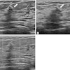
ABUS uses a high-frequency broadband transducer, a touch-screen monitor, and a dedicated workstation for 3D imaging analysis of the whole breast. While previous research shows that ABUS can differentiate between malignant and benign breast lesions, the researchers noted a lack of data on its use for assessing the efficacy of neoadjuvant chemotherapy.
CEUS meanwhile can help document changes in blood flow distribution within a tumor, which is more likely to occur at an early stage of chemotherapy. Along with that, it can help evaluate the architecture of blood vessels and microvessels.
Xie and colleagues wanted to evaluate the predictive accuracy of ABUS and CEUS, both individually and combined, for finding out treatment response after two cycles of chemotherapy in women diagnosed with locally advanced breast cancer.
The researchers collected data from 43 women with pathologically confirmed invasive breast cancer treated with neoadjuvant chemotherapy. The standard for evaluation they used for evaluating treatment response was based on surgery within 21 days of completing the treatment regimen.
The women underwent CEUS and ABUS one week before receiving chemotherapy and after two treatment cycles. For CEUS, the researchers explored rising time, time to peak, peak intensity, wash-in slope, and wash-in area under the curve for before and after chemotherapy. ABUS meanwhile measured maximum tumor diameters in the coronal and sagittal planes to calculate tumor volume. Images were analyzed by two radiologists with more than 10 years of breast ultrasound experience.
The team found after using logistic regression analysis that differences in tumor volume on ABUS, as well as time to peak and peak intensity on CEUS were independent predictors of complete pathologic response. The researchers also found that combining both ultrasound methods resulted in the highest diagnostic accuracy.
| Diagnostic performance for predicting treatment response | |||
| ABUS | CEUS | Combined method | |
| Area under the curve (AUC) | 0.891 | 0.918 | 0.95 |
The study authors wrote that while larger, multicenter trials are needed before wider implementation of the ABUS and CEUS combination, it could serve as an alternative to conventional ultrasound. They added it could also keep track of hemodynamic alterations within the tumor indicative of treatment response.
"Some studies proposed the use of computer-aided systems and artificial intelligence to reduce the observer variation in the determination of the region of interest on breast CEUS," the authors wrote. "Future studies should evaluate the role of these tools in improving the prediction of complete pathologic response following neoadjuvant chemotherapy."


