Endobronchial ultrasound may be used as a screening test for pulmonary hypertension in high-risk patients, a study published December 15 in JTCVS Techniques found.
Researchers led by Nathaniel Deboever, MD, from the University of Texas MD Anderson Cancer Center found that this ultrasound method achieved high performance and could be a superior alternative to transthoracic echocardiography for patients with pulmonary hypertension.
“Endobronchial ultrasound interrogation of pulmonary artery hemodynamics is safe and feasible,” Deboever and co-authors wrote.
Pulmonary hypertension is a physiologic variable in evaluating patients undergoing major thoracic operations. However, the researchers suggested that this is often neglected because of the need for right heart catheterization stemming from the inaccuracy of transthoracic echocardiography. While catheterization is considered a gold standard assessment method, the researchers wrote that “many” thoracic surgeons prefer avoiding this invasive method.
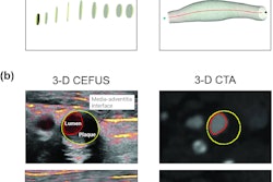
Endobronchial ultrasound is typically used in lung cancer patients as part of cancer staging. With this in mind, Deboever and colleagues studied the feasibility, value, and accuracy of an endobronchial ultrasound-based method for assessing pulmonary artery hemodynamics in patients scheduled for major pulmonary resection.
The team’s prospective study included 20 patients, all of whom also underwent right heart catheterization. The researchers compared the pulmonary artery pressure measurements taken by endobronchial ultrasound to the catheterization values. They reported that the prevalence of abnormal pulmonary artery pressure was 65% based on catheterization.
All patients underwent measurement of the pulmonary vascular acceleration time by endobronchial ultrasound with no adverse events, the researchers found. They also found through linear regression analysis a correlation between pulmonary vascular acceleration time and right heart catheterization (r = -0.059, p = 0.007).
When using a threshold of 140 milliseconds (msec) for the optimal pulmonary vascular acceleration time, the team reported an area under the curve (AUC) of 0.736 to predict pulmonary hypertension.
“This was used to calculate a positive and negative likelihood ratio following a positive diagnosis of 2.154 and 0.538 respectively,” the researchers wrote.
They added that using the 140-msec threshold had better sensitivity for predicting pulmonary hypertension than current guidelines of 105 msec used for transthoracic echocardiography.
The study authors suggested that in the future, endobronchial ultrasound could have utility for screening high-risk patients who undergo an extensive intrathoracic operation. However, they called for larger, multicenter studies to validate their results.
“Those with pulmonary hypertension on [ultrasound] can then be guided to have a right heart catheterization or potentially to measurement of the pulmonary artery pressure by direct puncture of the pulmonary artery, which has been shown to be safe in human and animal models with normal pulmonary pressures,” the authors wrote.
The full study can be found here.