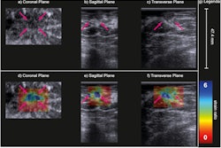
3D contrast-enhanced fusion ultrasound (CEFUS) can analyze carotid plaque volume burden and could monitor plaque development over time, a study published January 3 in Ultrasound in Medicine & Biology found.
Researchers led by Karin Yeung, MD, from the University of Copenhagen in Denmark found that this ultrasound method showed comparable performance to that of 3D CT angiography (CTA) and could provide visualization of the vessel lumen, creating a “lumenography.”
“3D CEFUS is promising for monitoring carotid plaque burden, with lumenography considered as an additional imaging biomarker,” Yeung and co-authors wrote.
Atherosclerotic plaque in the carotid artery is a risk factor for cardiovascular disease, with imaging used to monitor plaque development. 3D CEFUS is a novel technique that provides a spatial visualization of the carotid artery lumen. The resulting contrast-based “lumenography” resembles lumen visualization in angiography, the researchers highlighted.
Yeung and colleagues compared the respective performances of 3D CEFUS and 3D CTA in assessing carotid plaque volume. They acquired CEFUS images by spatial stitching of serial 2D images. To do this, they used an ultrasound system with magnetic tracking, a linear array transducer, and Bracco Diagnostic's SonoVue contrast agent.
The team also constructed atherosclerotic carotid arteries from both imaging modalities with lumenography using an offline software program. In total, the team included data from 39 carotid arteries.
 Segmentation of the carotid artery included the reconstruction of the carotid artery from 3D CEFUS/3D CTA to enable volume quantifications. (a) Only shown for 3D CEFUS, a 3D cylindrical model was initialized from manually set circle marks of the vessel wall border (media-adventitia interface) in the common carotid artery and internal carotid artery. From the circles, a mesh of the vessel border (yellow delineation) was formed. (b) The contour of the contrast-filled lumen was outlined (red delineation), creating a lumenography into the final reconstructed 3D carotid model. The same segmentation procedure was performed on the corresponding 3D CTA. The images are available to use under a Creative Commons license: CC BY 4.0 DEED, Attribution 4.0 International.
The average lumen and plaque volume in 3D CEFUS were 0.65 cm3 and 0.62 cm3, respectively, the researchers reported.
They also found that lumen volume differences between 3D CEFUS and 3D CTA did not achieve statistical significance. This included an average difference of 0.01 cm3 between the two p = 0.26), as well as a limits of agreement range of ±0.11 cm3. However, the plaque volume difference between the two modalities was -0.12 cm3 (p = 0.006) with a limits of agreement range of ±0.39 cm3.
The researchers also found good intrareader agreement of 3D CEFUS segmentations, including intraclass correlation coefficients (ICC) of 0.97 for lumen volume and 0.94 for plaque volume. Meanwhile, interreader agreement was also good with ICC values of 0.95 and 0.92 for CEFUS lumen and plaque volume, respectively.
Finally, the lumen was significantly smaller on the symptomatic side (0.54 cm3) than on the asymptomatic side (0.73 cm3, p < 0.05). Meanwhile, plaque volume was larger on the symptomatic side (82 cm3) than on the asymptomatic side (0.54 cm3, p < 0.05).
The study authors called for longitudinal studies on volumetric plaque burden development to validate 3D CEFUS. They also noted that the modality is currently limited by its reduced applicability, with only experienced sonographers being able to use the technology. They added that the modality calls for continuous ultrasound technology developments.
The full study can be found here.


