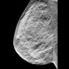
NEW ORLEANS - Cloning, colonization, and cannibalism are among the histological hallmarks of breast lesions that pathologists look for when analyzing specimens.
In a presentation at the Breast Imaging Conference on October 5, Dr. Whitaker Sewell, professor of pathology at Emory University in Atlanta, updated the 300 mammographers in attendance on the role of histological information in cancer management. The Medical College of Wisconsin and Instrumentarium Imaging, both of Milwaukee, are co-sponsors of the conference.Pathology begins with the normal anatomy of the breast. Investigators pay close attention to its epithelial structures, as well as to the terminal duct-lobular unit (TDLU), made up of the lobules, ductules, and terminal ducts.
"Most of the cancers originate in the TDLU, including cysts, fibroadenomas, and hyperplasia," Sewell said.
When trying to determine whether lesions are benign or malignant, pathologists study the epithelial structure closely. "All benign structures have both epithelial and myoepithelial cells. Once you have proliferation of only one type of cell -- epithelial -- that is the cancer," Sewell explained.
Colonizing comes into the picture when ductal carcinoma in situ (DCIS) is suspected. Advances in mammography techniques have greatly improved DCIS detection. As a result, pathologists have changed the way they classify DCIS, Sewell said. Rather than looking at the architecture of the lesion or even for the presence of necrosis, investigators study the size, uniformity, or irregularity of the nucleus.
For example, all types of DCIS have the capacity to conquer and colonize adjacent lobular and ductal structures. Low-grade DCIS, which generally forms cribriform and micropapillary patterns, has been shown to be particularly adept at invading other quadrants of the breast besides the ducts. Low-grade DCIS is also characterized histopathologically by a monoclonal population of cells with uniform characteristics.
In comparison, usual or ductal hyperplasia may have some of the same calcifications as low-grade DCIS, but these benign lesions will not be monoclonal. Instead, the pathologist will look for a mixture of cells that are derived from both the epithelial and myoepithelial areas of the duct wall.
Sewell highlighted other histological signposts of benign, nonproliferative disorders of the breast including:
- Duct ectasia. This secretory disease involves ducts and is associated with periductal inflammation. On a mammogram, large, rod-like calcifications are often seen with these lesions.
- Phyllodes tumors. Similar to fibroadenomas, these leaf-shaped tumors consist of a mixture of stroma and epithelial structures.
"Phyllodes tumors are remarkable in that they tend to occur in older women, grow faster, and have a more irregular periphery," Sewell said. Narrow excision of these tumors can leave some of the peripheral extensions behind, increasing the potential for recurrence.
In the hunt for invasive ductal carcinoma, pathologists focus on growth patterns such as irregular-mass lesions and spiculated borders. One type of invasive ductal carcinoma is tubular carcinoma. On a mammogram, the density of these lesions makes them fairly easy to detect, Sewell said. In addition, tubular carcinoma is generally associated with low-grade DCIS.
"High-grade DCIS and tubular carcinoma don't go together," he said. "If someone says that they think it's tubular carcinoma with high-grade DCIS, make them check again."
Finally, some of the trickiest benign, proliferative lesions for pathologists to study are radial scars. The process that causes radial scars is unknown, Sewell said, although chronic ischemia or infarction are two possibilities. The spiculated appearance of a scar may mimic a lesion. The aim of the histological exam is to distinguish the ductules trapped inside the scar and rule out infiltrating carcinoma.
What Sewell termed "cannibalism" comes about when radial scars are present along with other lesions. According to a 1999 study in the New England Journal of Medicine (Feb 11, 1999, 340:6), a patient with usual hyperplasia and radial scar formation could be three times as likely to develop cancer than a patient with only the benign lesion. In addition, the study demonstrated that the risk could increase as the number and size of the scars increase.
While researchers have yet to determine the pathologic process that causes radial scars or whether the lesions actually alter the risk of breast cancer in women with benign disease, pathologists do not believe that benign radial scars can transform into carcinoma, Sewell said.
By Shalmali Pal
AuntMinnie.com staff writer
October 6, 2000
Let AuntMinnie.com know what you think about this story.
Copyright © 2000 AuntMinnie.com


