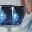
Changes in breast density can affect the performance of computer-aided detection (CAD) software used to analyze mammography images, according to results of a study from Spain presented at the European Congress of Radiology (ECR).
The researchers found that CAD's performance varied by breast density, with the software performing better in women with fatty breasts as opposed to those with dense breasts. Results of the study were presented at ECR by Dr. Cristina Romero of Hospital Virgen de la Salud in Toledo.
The researchers tested a commercially available CAD algorithm (ImageChecker version 5.4, Hologic, Bedford, MA) on 8,750 mammograms acquired from January to December 2007 on a full-field digital mammography (FFDM) machine (NovationDR, Siemens Healthcare, Erlangen, Germany).
Images were read by two experienced breast radiologists and rated using BI-RADS criteria for lesion assessment (BI-RADS 1-5) and breast composition (BI-RADS 1 = almost entirely fat, 2 = scattered fibroglandular densities, 3 = heterogeneously dense, and 4 = extremely dense).
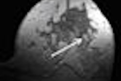
Of the 8,750 cases, 138 were cancers, with the CAD software correctly marking 118 for a sensitivity of 86.5% and a false-positive rate of less than two marks per case. In addition to density, CAD's effectiveness in different breast tissue types varied based on the type of lesion: CAD performed well in finding microcalcifications regardless of tissue type, but the software's accuracy varied widely for masses and other types of architectural distortion.
CAD performance by lesion type and breast density
|
The difference in CAD's performance for microcalcifications was not statistically significant, Romero said, but it was significant for masses, asymmetry, and architectural distortion.
In discussing the group's results, Romero cited previous research by Yaffe et al that hypothesized that CAD could eventually be used in a prescreening role to review some low-risk cases prior to a radiologist's review, with radiologists only reading cases that had CAD marks.
"Our results support this idea when the CAD is used under certain conditions," Romero said. "It may be possible to do prescreening with CAD in BI-RADS 1 and 2 density cases."
More research is needed before CAD can be used in this kind of environment, she added. For example, the researchers are pursuing a two-year follow-up study to examine the impact of CAD false negatives.
By Brian Casey
AuntMinnie.com staff writer
March 25, 2009
Related Reading
CAD boosts FFDM performance, March 4, 2009
Breast MRI CAD algorithm predicts cancer metastasis, February 6, 2009
CAD can boost double-reading detection rate, January 22, 2009
Interactive use of mammo CAD may improve mass detection, December 22, 2008
Breast MRI pinpoints residual disease after chemotherapy, December 19, 2008
Copyright © 2009 AuntMinnie.com

