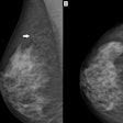
Standard image processing algorithms help mammography detect calcification clusters more effectively than low-contrast or film-screen algorithms and could reduce false positives, according to a new study published in the August issue of the American Journal of Roentgenology.
"For calcification clusters, there was a significant decrease in the number of false positives reported on normal images for standard image processing compared with film-screen image processing," wrote lead author Dr. Lucy Warren, of the National Coordinating Centre for the Physics of Mammography at Royal Surrey County Hospital in Guildford, U.K., and colleagues.
Every manufacturer applies its own image processing algorithm to breast images, which produces varied image results. Although studies have been performed to explore the effects of image processing on mammography, they have not been analogous, Warren's group wrote.
"Different image processing algorithms were used on systems with different detectors, different radiologists inspected different images, localization was not included in the task, not all cases were from different patients to maintain the independence assumed in the analysis, the cancers tended to be obvious and easily detected in all image processing types investigated, and only calcifications were investigated," they wrote.
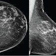
The researchers designed the study to address these limitations. Data include 270 pairs of breast images (both breasts, one view) collected from eight Hologic systems with amorphous selenium digital detectors. Eighty image pairs showed breasts with subtle malignant masses, 30 image pairs had biopsy-proven benign lesions, 80 image pairs had simulated calcification clusters, and 80 image pairs had no cancer (AJR, August 2014, Vol. 203:2, pp. 387-393).
Warren and colleagues ran the image pairs through three types of Hologic image processing:
- Standard (full enhancement)
- Low contrast (intermediate enhancement)
- Pseudo film-screen (no enhancement)
Although the low-contrast and film-screen image processing algorithms were developed by Hologic for investigational purposes only, the researchers included them to represent the wide range in image processing algorithms seen on the different digital mammography systems currently used in clinical practice.
Seven experienced mammography readers (six radiologists and one radiographer) interpreted the images, identifying and rating regions they suspected to be cancerous for likelihood of malignancy. The researchers then analyzed the results using the jackknife free-response receiver operator characteristics (JAFROC) method.
Reader-averaged JAFROC figures of merit (FOMs) for each image processing algorithm are shown in the table.
| FOM ratings by image processing type | ||
| Image processing algorithm | Noncalcification cancers | Calcification cancers |
| Standard | 0.726 | 0.652 |
| Low contrast | 0.717 | 0.628 |
| Film-screen | 0.719 | 0.612 |
"Our work found that the standard image processing algorithm provided significantly higher detection of calcification clusters than the low-contrast and film-screen image processing algorithms," Warren and colleagues wrote. "[We] also found that there were no significant differences in detection of noncalcification cancers between the different types of image processing."
The study also showed that using the standard image processing algorithm produced fewer false positives, according to the authors. The number of marks for false-positive calcifications increased by 15% for film-screen image processing compared with the standard algorithm.
The study results are promising, but they only apply to the particular image processing algorithms used, the researchers cautioned.
"We recommend that objective measurements, such as the method used in this study, be applied when selecting the optimal image processing to be used by radiologists with other image processing algorithms and systems," they wrote.


