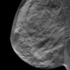
Women receive about 30% less radiation during screening mammography than has long been assumed, which suggests that the "harm" of radiation dose in mammography also has been overestimated, according to research presented on July 15 at the 2015 American Association of Physicists in Medicine (AAPM) meeting in Anaheim, CA.
By correcting misinformation about radiation dose in mammography, the study findings could influence the ongoing controversy over when and how often women should undergo breast cancer screening, said study presenter Andrew Hernandez in a press conference held by AAPM. Hernandez is a doctoral candidate at the University of California, Davis (UCD) in the lab of John Boone, PhD.
 Andrew Hernandez from the University of California, Davis.
"Our findings are consistent with other studies that have shown that, historically, radiation dose in mammography has been overestimated by 30%," Hernandez said. "This means that for the average patient, this particular risk associated with mammography has also been overestimated by 30%."
Heterogeneous, not homogenous
The breast is composed of skin, fat, and glandular tissue, with glandular tissue having the greatest risk of radiation damage, according to Hernandez and colleagues. Conventional methods of estimating radiation dose in mammography have assumed that the breast is a uniform mixture of glandular and fatty tissue. Instead, the distribution of the two types of tissue can vary widely.
"Three-dimensional imaging techniques can help us understand how glandular and fatty tissue is actually oriented in the breast, and we can use this information to estimate the dose impact," Hernandez said.
To determine how glandular tissue is distributed throughout the breast, Hernandez and colleagues used a model of breast anatomy based on the CT exams of 219 women who represented a large population average and ranged in age, ethnicity, and breast size and density.
When the disparate nature of breast tissue is taken into account, mammography's radiation dose is overestimated by 30% on average, the group found. And because the amount and distribution of glandular tissue varies from woman to woman, some could be receiving 20% less radiation, while others could receive 40% less.
In any case, the study data suggest that the harms of mammography are smaller than they've been perceived -- which means that mammography's efficacy is greater, Hernandez said. However, further work is needed to make dose estimates more accurate before the study findings can be applied to clinical practice.
"Current methods for radiation dose estimations in mammography need to be fundamentally updated to address the realistic distribution of glandular tissue within the breast," he concluded.


