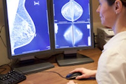
Women who undergo mammography screening at breast centers with high volumes tend to have better outcomes, which is encouraging news for a modality often criticized for its tendency to overdiagnose. Study results were published online August 11 in the Journal of Medical Screening.
Mammography quality can be measured by characteristics of the images, the proportion of women called back for additional imaging, sensitivity, specificity, and positive predictive value, wrote the group led by Tracy Onega, PhD, of Dartmouth College. But it's really the detection of small, early-stage tumors that haven't spread to the lymph nodes that influences mortality rates.
"It is only by diagnosing and treating invasive cancers early, and thereby preventing progression to more advanced disease, that mammography can achieve mortality benefit," Onega and colleagues wrote (J Med Screen, August 11, 2015).
The right cancers
Although plenty of research has been published on variation in breast screening performance by physician volume, less has been published on facility volume, according to the researchers.
"Measuring facility interpretive volume may be more practical ... because many radiologists practice at more than one facility, making accurate tracking across multiple facilities challenging," they wrote.
 Tracy Onega, PhD, from Dartmouth College.
For the study, the researchers calculated annual mammography interpretive volumes from 2000 to 2009 for 116 facilities in the U.S. Breast Cancer Surveillance Consortium (BCSC) network, using radiology, pathology, cancer registry, and women's self-reported information to identify the indication for each exam and cancer and patient characteristics.
The group then explored the effect of annual total volume and percentage of screening mammograms on cancer detection rates. Tumors were defined as having a "good prognosis" if they were screen-detected invasive cancers smaller than 15 mm, early-stage, and lymph-node-negative at diagnosis.
Out of 3.1 million screening mammograms conducted during the study period, about 10,000 cancers were screen-detected within one year of a woman's initial exam. The majority of facilities (80%) had annual total interpretive volumes of more than 2,000 mammograms; 42% had volumes of more than 5,000 and almost 20% had volumes of more than 10,000.
Onega and colleagues found that higher volumes translated into better prognoses. Those facilities that read more screening mammograms were significantly more likely to diagnose invasive tumors with good prognoses. The researchers also observed an accompanying decrease in tumors with poor prognoses. In fact, compared with facilities with an average volume of 1,000 to 2,000 mammograms interpreted per year, those with an annual average volume of 5,000 to 10,000 per year were 32% more likely to diagnose a "good prognosis" cancer.
"Our results suggest better early invasive cancer detection above 2,000 mammograms interpreted annually at a facility, which is consistent with ... prior findings suggesting improved screening performance with 2,000 exams interpreted annually for radiologists," the authors wrote.
And the findings seem to underscore the idea that more volume doesn't necessarily lead to overdiagnosis, Onega noted.
"We want screening mammography to reduce mortality -- and to avoid overdiagnosis," she told AuntMinnie.com. "It looks like facilities that interpret more mammograms have a higher likelihood of finding more 'good prognosis' cancers."
So how might these findings translate into clinical practice? There appears to be a relationship between facility volume and finding invasive cancers early, Onega said.
"Our study suggests that there may be volume benchmarks, and a low-volume facility may expect better outcomes if they bump up the number of mammograms read per year," she said.


