Wednesday, November 29 | 10:40 a.m.-10:50 a.m. | SSK02-02 | Room E451A
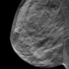
A computer system based on deep-learning technology was as efficient as radiologists in detecting breast cancer on digital mammography exams in a study by a group from Sweden and the Netherlands.Six radiologists with a combined experience of 22 years in digital mammography retrospectively looked at 155 unilateral two-view exams, which included 73 malignant and 82 negative results. Among the 82 negative results, 42 masses were biopsy-proven as benign, while 40 normal cases were ruled as BI-RADS 1 or 2 with one-year follow-up. All lesions were rated for suspiciousness on a scale from 1 to 10, with the highest score used as the overall exam rating.
The researchers then used a commercially available algorithm based on deep-learning technology (Transpara 1.2.0, ScreenPoint Medical) to analyze the dataset to automatically identify soft-tissue lesions and calcifications. The method combines the findings of all available views into a single cancer suspiciousness score of 0 to 10.
The group found no statistically significant difference between the performance of the system and any of the six readers, though the area under the curve was slightly higher for the radiologists.
"These encouraging results point to the possibility of implementing artificial intelligence-based reading of mammograms for population-based breast cancer screening programs," said presenter Alejandro Rodriguez-Ruiz from the department of radiology at Radboud University Medical Centre in Nijmegen, the Netherlands. "A computer system such as this one could be used, for instance, to automatically discard cases which are most likely normal, reducing the workload and leaving to the radiologists a case set to be read with a higher incidence of cancer."


