Wednesday, November 29 | 11:40 a.m.-11:50 a.m. | SSK02-08 | Room E451A
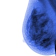
In this session, researchers will present a machine-learning method for analyzing parenchymal patterns from digital mammography screening to assess breast cancer risk and how it may change over time.Led by Jun Wei, PhD, an associate research scientist at the University of Michigan in Ann Arbor, the researchers are hopeful that the imaging technique could lead to a personalized screening regimen for patients and early cancer detection.
The retrospective study featured data from 398 patients and 199 paired cancer cases, as well as a cohort of cancer-free control subjects who were matched for years of screening, age, and race. For each subject, the researchers collected screening digital mammograms from the year of the cancer diagnosis and up to five years of consecutive prior screening.
In all, they analyzed more than 2,700 craniocaudal views, with three regions of interest automatically localized and used for mammographic parenchymal pattern analysis.
The mammographic parenchymal pattern could be used to differentiate the patients from the matched controls, Wei and colleagues found. In addition, the changes in mammographic parenchymal patterns increased as the time of cancer diagnosis approached.
"Future work is underway to enlarge the dataset and to improve the machine-learning scheme," the group wrote.


