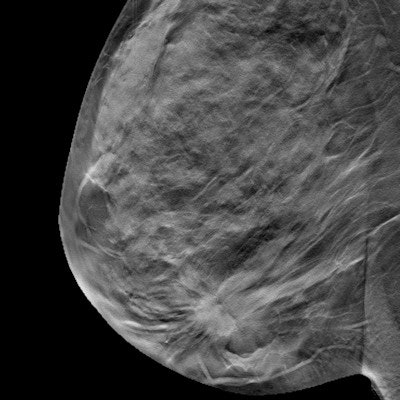
It's not just for screening anymore: A new study found that using digital breast tomosynthesis (DBT) in the diagnostic setting reveals more cancers and produces fewer false positives than 2D digital mammography, suggesting that DBT's proven advantage for breast screening also carries over into the diagnostic realm.
The results are good news because a lower false-positive rate in the diagnostic setting can lead to improved patient care, said presenter Dr. Charmi Vijapura from Massachusetts General Hospital in a talk at the recent American Roentgen Ray Society (ARRS) meeting in Washington, DC.
"Our results show that using DBT can positively affect patient care by reducing unnecessary biopsies -- while also maintaining a high cancer detection rate," she said.
Diagnostic demand
DBT's benefits have been studied primarily in the screening setting, Vijapura said. The impact of DBT on false positives in the diagnostic setting has not been well-established. To address this lack of data, the researchers compared false-positive rates between diagnostic 2D mammography and DBT exams; they then compared the types of mammographic findings that led to false positives between the two modalities.
Vijapura and colleagues reviewed 22,324 2D diagnostic mammograms performed between August 2008 and February 2011 (before the hospital integrated DBT into its practice) and 23,446 diagnostic DBT exams performed between January 2013 and July 2015 (after DBT was integrated). Women who underwent these exams did so upon recommendation for short-interval follow-up, further evaluation of a breast problem, or following a concerning finding on MRI, CT, or PET/CT. Patients recalled from screening were not included in the study.
"We searched our mammography information system for all diagnostic mammogram reports and collected pathology results from within one year after the diagnostic mammogram was performed," Vijapura said. "We also culled data from tumor registries from five hospitals within our healthcare system."
The researchers considered a mammogram to be a false positive if there was no diagnosis of breast cancer within one year after tissue diagnosis or surgical consultation had been recommended (based on initial exams categorized as BI-RADS 4 or 5).
The group found that 3D mammography identified more cancers and produced fewer false positives than 2D mammography in the diagnostic setting.
| 2D versus 3D mammography for diagnostic use | |||
| 2D diagnostic mammography | 3D diagnostic mammography | p-value | |
| Cancer detection rate (per 1,000 exams) | 31.3 | 38.2 | p < 0.01 |
| False-positive rate | 7.3% | 6.5% | p < 0.01 |
"The false-positive rate decreased after DBT was integrated into the diagnostic setting, which suggests that the technology could improve the selection of patients recommended for biopsy," Vijapura noted.
In terms of which mammographic features prompted false positives, architectural distortion and masses led to more false-positive examinations on DBT than on 2D mammography, at 5% compared with 1% for architectural distortion and 33% compared with 20% for masses, the group found.
| Findings leading to false-positive exams on diagnostic mammography | ||
| 2D diagnostic mammography | 3D diagnostic mammography | |
| Architectural distortion | 1% | 5% |
| Asymmetry | 18% | 12% |
| Calcifications | 37% | 27% |
| Masses | 20% | 33% |
| Other | 24% | 23% |
Consider carefully
DBT reduced the number of false positives associated with asymmetries and calcifications, but the data regarding architectural distortion and masses are worth considering, Vijapura said.
"The types of findings that led to false-positive exams after DBT was integrated at our facility provide insight into the advantages and disadvantages of this new imaging modality," she said. "For example, a recent study found that architectural distortion is more readily detected on DBT than digital mammography but is less likely to be malignant on DBT than on 2D -- which could make biopsy recommendations more effective."


