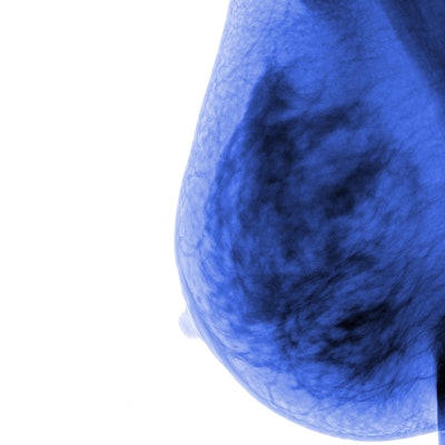
Interval cancers show less-favorable molecular features compared to cancers detected on screening mammography, making earlier diagnosis even more important, a study published February 3 in Academic Radiology found.
Researchers led by Dr. Emily Ambinder from Johns Hopkins Medicine in Baltimore found that interval cancers more often have higher tumor and nodal staging, are more often triple-negative, and have high Ki67 proliferation indices. They also found that interval cancers are more associated with dense breast tissue.
"The results of this study demonstrate the need for additional and/or novel supplemental screening techniques," Ambinder and colleagues wrote.
Screening mammography is the gold standard for detecting breast cancer in women. However, some women are still diagnosed with breast cancer between annual mammograms, known as interval cancers. This is due in part to mammography missing some breast cancers and producing false-negative reports. Lower breast density, no family history of breast cancer, and screening with digital breast tomosynthesis (DBT) have been reported to have lower associations with interval cancers.
In hopes of better informing and improving breast cancer screening for women, Ambinder and colleagues wanted to identify characteristics in patients, imaging, and tumors that differentiate screen-detected breast cancers from interval breast cancers.
The researchers looked at data collected between 2013 and 2020 from a total of 1,232 patients with an average of 64. Patient age, race, and personal history of breast cancer were similar between the screen-detected and interval cancer groups.
They found that the sensitivity of screening mammography was 92%, including 1,136 screen-detected cancers and 96 interval cancers. However, 21% (20/96) of patients had a high-risk screening breast MRI in between annual screening mammograms that identified the interval breast cancer.
The team also found that interval cancers were more linked to more unfavorable characteristics such as tumor staging, nodal staging, and triple-negative status, among others.
| Prevalence of screen-detected, interval breast cancer characteristics among patients | |||
| Screen-detected | Interval | p-value | |
| Primary tumor stage II or higher | 12% | 43% | < 0.001 |
| Regional lymph node stage I or higher | 12% | 22% | 0.003 |
| Triple-negative status | 6% | 21% | < 0.001 |
| High Ki67 proliferation indices | 38% | 62% | 0.002 |
Finally, patients with interval cancers more often had dense breast tissue. This included 75 out of 96 cancers (78%) in the interval group and 694 out of 1,136 (61%, p < 0.001). A family history of breast cancer in a first-degree relative was also associated with a higher likelihood of being diagnosed with an interval cancer.
The researchers called for future studies to include strategies to modify breast density in older women. They wrote that this could improve screening mammography's sensitivity.
The team found no significant links between having an interval cancer and whether the index screening mammogram was performed with full-field digital mammography or DBT.
The study authors also advocated for supplemental screening or alternative methods such as contrast-enhanced MRI, abbreviated MRI, ultrasound, and liquid biopsy.
"Although our current mammographic screening recommendations have a high sensitivity for breast cancer, there are women who are underserved and may benefit from the addition of other screening methods or more frequent screening," the authors wrote. "Further evaluation is needed to determine the optimal screening algorithms for all women."


