By offering interventional radiology trainees what they need most -- practice -- virtual reality training may be poised to stand in for the conventional version, according to a recent talk by researchers who have developed an immersive virtual reality training environment.
A team from Massachusetts General Hospital and Harvard Medical School has built a virtual reality (VR) production process to develop training modules that are being deployed to residents and medical students for training in interventional radiology (IR) procedures. A large majority of students are finding the sessions helpful and believe they have a future, according to surveys gauging the results.
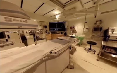 2D rendition of a 3D VR screenshot inside an interventional suite. Image courtesy of Dr. Colin McCarthy.
"The thing with any procedure-based specialty like interventional radiology is that a lot of it is practice and repetition. The subject is sometimes difficult, and there's a learning curve," said Dr. Colin McCarthy in a presentation at the Society for Imaging Informatics in Medicine (SIIM) 2015 meeting in May in National Harbor, MD. "[With VR] you build a suite of tutorials ... that allow generally novice people who haven't done procedures before the opportunity to work to get a feel for what's involved."
Advanced viewer, homemade video capture
Virtual reality isn't new. Since the stereoscopes of the mid-1850s, it has leveraged the slightly different views created by the separation of a person's two eyes to capture the world in 3D.
"Each eye gets a separate image, then your brain does the rest, giving you the perception of depth," McCarthy said. Interest in VR "has kind of waxed and waned over the years, but it's seen a resurgence recently because of head-mounted displays."
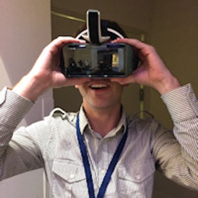
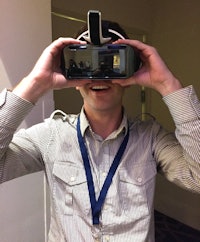 A SIIM attendee (and later, a reporter) were impressed by a high-resolution 360° view of an operating theater inside the Oculus Rift viewer.
The displays certainly have advanced. In the Mass General/Harvard project, McCarthy and colleagues Dr. Alvin Yu, Synho Do, PhD, Dr. Steven Dawson, and Dr. Raul Uppot built an immersive virtual reality experience using the Oculus Rift 360° viewer, a kind of high-tech viewing helmet originally designed as a gaming system.
The Oculus Rift combines a low-latency gyroscope with two low-persistence OLED displays placed directly in front of the viewer's eyes that block out external views. When the viewer rotates his or her head horizontally or vertically, Oculus tracks the movement and displays the appropriate image to simulate a 360° representation of the surrounding environment.
For video capture, the researchers tied together GoPro stereoscopic cameras to create a continuous view in a 360° space, and then delivered the finished product to medical students and residents for testing, McCarthy said.
"We targeted it at residents, new fellows, and medical students, and reviewed [interventional radiology] procedures -- some of the more basic things like biopsy -- and then reviewed safety topics like how to manage a medical emergency," he said.
Two video-capturing configurations were built to shoot the tutorials. The first is a stereoscopic 3D system that uses two GoPro Dual stereoscopic cameras, slightly set apart. The images are fused by software to deliver a single side-by-side elongated image.
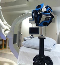 Seven GoPro Dual cameras mounted on a stand capture a high-definition, 360° view of the IR suite. Image courtesy of Dr. Colin McCarthy.
The second, a stereoscopic 360° camera, consists of seven of the GoPro cameras mounted in all directions on an aircraft-grade nylon stand. This setup captures the space entirely from the center of the room, according to McCarthy.
"We see the potential for this mainly as an introduction to a procedure room, but also if there's a contrast reaction or medical emergency, you can see people running in and out of the room," he said.
Each GoPro Dual produces its own video feed, which is stitched digitally to the output from the other cameras. The investigators draw vertical lines to set the horizon straight, affording the viewer a seamless and level circle from which to view the scene.
Video editing software (Adobe Premiere) is used for advanced editing. It also handles simple tasks such as blurring out patients' faces and drawing boxes to mask patient ID labels. The radiologists then added CT images and a voiceover for the various components before the content was distributed.
The group created several training modules for initial testing, including the following:
- Introduction to the interventional radiology suite
- How to set up a basic IR tray
- How to perform a CT-guided biopsy, femoral puncture, and angiogram
- How to treat a bleeding hypotensive patient
Early experiences
For initial testing, 12 participants were included: six attending radiologists, five fellows, and one resident.
After completing the module on setting up an IR tray, the participants rated the quality of their training experience via an online survey, which also tested their knowledge of the content and their ability to perform the tasks learned in the session. Nine (75%) of the 12 surveyed rated the module as "good" or "excellent."
When asked how they had previously trained for interventional radiology, about half said they read books and 55% attended lectures. Sixty-eight percent reviewed literature, attended conferences, or received hands-on supervision. Only one-third had ever used virtual reality training.
Fifty-eight percent said the VR headset would definitely help their IR training in the future, and the remaining 42% said it might help.
"We did get useful comments; a lot of people described motion sickness, which is very well-described in virtual reality literature," McCarthy said. "After a while you get used to it, but at first it's very disorienting."
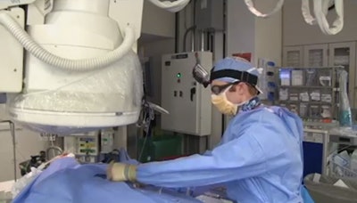 Gastrostomy procedure conducted as part of a VR session. Image courtesy of Dr. Colin McCarthy.
The virtual future
Going forward, the group would like to integrate a Leap Motion controller, which senses the movements of hands and fingers and follows every gesture. Also on the wish list is building a virtual reality studio, which would allow the team to create a VR environment and walk around in it, rather than being "stuck in the middle of the room" with the 360° camera. The graphics could also use some improvement, and even haptic interaction isn't out of the question.
"We'd like to get haptic feedback using gloves, which is completely separate and much more advanced, but it's what people suggested to us," he said.
There is certainly precedent for effective virtual reality training. A 2012 Swedish study discussed a VR module used to train surgeons in performing cholecystectomy; researchers found that participants in VR training made significantly fewer errors than nonparticipants.
McCarthy was skeptical of the value of VR when he first heard of it, but no more.
"Now I see it as something that in the future will be potentially useful as an adjunct to conventional training," he said. "The question is the cost and whether you could deploy this to a group of medical students. We think it has tremendous potential."


