Dear Advanced Visualization Insider,
Interventional radiology is a subspecialty that requires high skill levels, painstaking control of movement, and plenty of practice. So what better way to teach trainees the skills they'll need than by teaching them in virtual reality, where there's no limit on how often they can start the lesson over again?
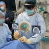
Such was the idea behind Dr. Colin McCarthy's project at Massachusetts General Hospital (MGH) to create a virtual reality production process to teach some of the key skills interventionalists need, such as for biopsies, cholecystectomies, and even just setting up the work space.
Armed with arrays of stereoscopic cameras and a 360° viewer originally designed for gaming, the group has created a series of virtual reality training modules in high-resolution videos. How do the trainees feel about learning this way? Find out in our Insider Exclusive.
There was a time when color in medical imaging was an afterthought, but now it's becoming increasingly important in a wide range of advanced imaging applications. Color has never been standardized in the same manner as grayscale imaging, so a hue can denote one thing in one application and quite another in another.
The International Color Consortium has standardized color in several industries, but medical imaging needs more accuracy. What's happening in medical imaging? Find out here.
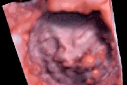
Color is also an important component of a new application aiming to provide realism in echocardiography. An advanced algorithm looks for the best image pixels on which to base its full-motion 4D color videos of the human heart. Get the rest of the story by clicking here.
Parathyroid lesions fall into three distinct types that 4D CT can now distinguish according to their signature enhancement patterns, according to a new study from Duke University Medical Center. Three phases are needed to distinguish all of the patterns, but it's a small price to pay for the correct diagnosis, researchers say.
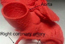
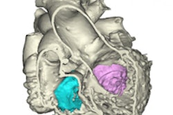
There's a new and cost-efficient way to print heart components for surgical planning and education, according to another study from MGH researchers. Our how-to article shows just how they did it. Not to be outdone, a Grand Rapids, MI, group's heart printer makes use of both echo and CT.
Finally, radiologist performance with computer-aided detection of lung nodules comes with so many variables that optimizing detection might seem impossible, according to a talk at the International Symposium on Multidetector-Row CT. But several factors have been identified that make a big difference in how well radiologists detect the most important findings. Learn what matters here.
You'll find the latest on radiology's newest ways of seeing things right here in your Advanced Visualization Community.


