Dear Advanced Visualization Insider,
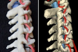
In the blink of an eye -- or so it seems -- 3D printing has progressed from hobby status to one of radiology's hottest topics. Perhaps this is due to the promise of a vast array of clinical applications made possible by the technology, from medical models to implants and beyond.
3D printing was the focus of a March 31 webinar presented by the Society for Imaging Informatics in Medicine (SIIM). In the hour-long session, three radiologists from the University of Utah shared their experiences with the new 3D printing lab they set up a year ago, talking about the products and possibilities the technology has created and the software that makes it all run.
Part 1 of our two-part series covers the hardware and history of 3D printing, explores some clinical applications, and even looks at the gooey, messy side of printing technologies. In part 2, we explore the software behind the discipline, some lessons learned, and why radiologists are an essential part of the process that can't be replaced. You'll find part 1 here, and part 2, brought to you before our other members can access it, is our Insider Exclusive. Perhaps their success will inspire you to create some 3D printing stories of your own.
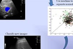
In the field of computer-aided detection (CAD), researchers from Rhode Island have created a versatile algorithm that can diagnose several diseases at once using quantitative texture analysis, according to an article in the Journal of Medical Imaging. Relying on quantitative ultrasound, the Brown University team is exploring three conditions that have been getting a lot of attention lately: craniosynostosis, steatosis, and adenomyosis. Find out how they did it, and what they might look at next, by clicking here.
Also, read about an intriguing study from Canada in which researchers used CAD as part of a CT lung cancer screening program to help nonradiology personnel triage which scans should be passed on to radiologists for further review. Find out how the program worked by clicking here.
In Ohio, researchers used analytics software to determine which estrogen-receptor-positive women with breast cancer actually need chemotherapy and which have nonaggressive cancers that require less intensive treatment. The test uses textural kinetics analysis, which measures dynamic changes in breast lesion texture during contrast uptake, for cancers considered to be low-risk. If successful, it could go a long way toward reducing costs and unnecessary treatment for women with breast cancer, according to our story by Senior Editor Erik L. Ridley.
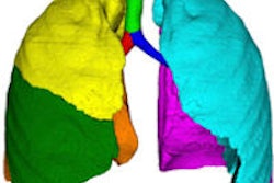
Finally, a new algorithm can be used to assess the extent of airway disease, according to researchers from Germany, who developed a lobe-based segmentation method to evaluate children with cystic fibrosis. Learn how they did it here.
We invite you to scan through the links below for the rest of the news in 3D and advanced visualization, served up fresh and hot in your Advanced Visualization Community.


