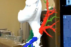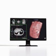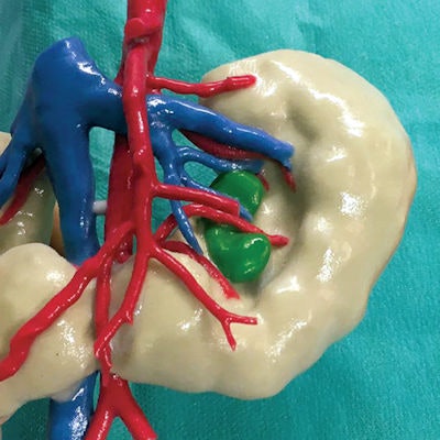
Augmented reality (AR) and 3D printing provided better visualization of intricate kidney structures than conventional MRI and CT -- improving physicians' understanding of patient anatomy before performing kidney tumor surgery, according to an article published online April 19 in JAMA Network Open.
Researchers from the Netherlands created personalized 3D-printed and augmented reality 3D models of the kidneys of pediatric patients with Wilms tumors, the most common kidney cancer in children. Clinicians who examined these 3D models found that they helped pinpoint the location of kidney tumors and relevant anatomical structures during preoperative planning more precisely than standard techniques.
"New, preoperative 3D imaging processing strategies helped increase the surgeons' knowledge about the kidney anatomy, thus assisting in improving the planning of tumor resection and optimizing the procedure of choice: nephron-sparing surgery or nephrectomy," first author Dr. Lianne Wellens from Princess Máxima Center for Pediatric Oncology and colleagues wrote. "This method could improve radicality of tumor resection and spare healthy kidney tissue, thereby preserving long-term renal function."
Innovative supplementary visualization
Traditionally, clinicians have relied on conventional imaging modalities to diagnose kidney tumors and to plan their surgical approach. Although the survival rate of patients with Wilms tumors who undergo treatment is high (between 80% and 90%, depending on the severity of the tumor), risks of long-term kidney function loss and perioperative complications still remain.
The recent emergence of advanced visualization techniques such as 3D printing and AR has opened up new avenues for examining complex anatomy that may enhance clinicians' ability to locate key structures in and around the kidneys of patients with Wilms tumors, the authors noted. These techniques can facilitate personalized surgical planning -- potentially increasing the success of tumor resection and minimizing the need for reoperation and additional radiotherapy.
To test this notion, Wellens and colleagues acquired the MRI and/or CT scans of 10 children diagnosed with Wilms tumors between January 2016 and May 2017. The average age of the patients was 3.7 years, and 70% were girls.
Next, they segmented the kidney parenchyma, tumor, arteries, veins, and urinary collecting ducts from the scans using segmentation software (Mimics Innovation Suite, Materialise) and then saved the images in stereolithography (STL) format. They sent these virtual 3D models to Materialise, where specialists used them to create multicolor 3D-printed kidneys with a 3D printer (ZPrinter, Z Corporation).
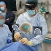
The researchers also exported the virtual 3D models into 3D software (Meshmixer, AutoDesk) to optimize them for viewing with a wireless AR headset (HoloLens, Microsoft). The AR head-mounted display allowed users to see and interact with the 3D kidney models via hand gestures and voice instructions.
Finally, a panel of seven pediatric physicians completed a survey comparing the utility of the different imaging methods on a five-point Likert scale. Their assessment included ratings of the visibility of key anatomical structures as well as the extent to which the different methods facilitated presurgical planning and decision-making regarding treatment.
Future of preoperative planning
Overall, the AR and 3D-printed models received higher scores than conventional MRI and CT for the depiction of four key anatomical structures: the tumor, arteries, veins, and urinary collecting ducts. (A score of 5 indicates best visibility, and 1 indicates worst visibility.) The differences in scores were statistically significant for all structures between conventional imaging and 3D printing or AR but never between the two 3D visualization techniques themselves.
| Visualization of kidney structures using different imaging techniques | |||
| Anatomical structure | MRI, CT | 3D printing | AR |
| Tumor | 4.07 | 4.67 | 4.71 |
| Arteries | 3.62 | 4.54 | 4.83 |
| Veins | 3.46 | 4.5 | 4.83 |
| Urinary collecting ducts | 2.76 | 3.86 | 4 |
The 3D models were useful not only for the visualization of key kidney structures but also for studying ideal tumor access points and possible complications before performing surgery, Wellens and colleagues noted.
"The novel 3D visualization techniques presented here may be a useful added tool when planning nephron-sparing surgery in unilateral and bilateral [Wilms tumors]," they wrote.
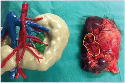 Kidney specimen (right) with corresponding 3D-printed kidney (left). The Wilms tumor is outlined in yellow in the kidney specimen and colored green in the 3D-printed model. Image courtesy of Wellens et al. Licensed under CC BY.
One of the main reasons why the new techniques provided better visualization of the kidney than conventional imaging was that the 3D models allowed the physicians to view inside the kidney. The 3D-printed kidneys had small windows that revealed a full view of the tumor and its border with healthy tissue and vasculature. With AR, users were additionally able to rotate, adjust, and manipulate transparent 3D structures.
Augmented reality and 3D printing have notable limitations in this application, however, such as high costs and long production times. It cost roughly $500 and took up to five days to manufacture each multicolored 3D-printed kidney. And though the group was able to generate the AR 3D models in less than two hours, an AR headset typically costs upwards of $3,000.
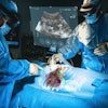
Yet the potential for these technologies to improve surgical outcomes and preserve long-term renal function may outweigh these costs, the authors noted. This may be particularly relevant for complex cases. In one challenging case, for example, the tumor of one of the pediatric patients was encased inside a horseshoe kidney. The unusual structure of the kidney prompted the surgical team to refer to the 3D-printed model for real-time guidance during surgery.
For this situation, the 3D-printed kidney had "significant added value" compared with conventional imaging by facilitating intraoperative guidance, they wrote. Thus, the technique "may be considered an innovative supplementary visualization in clinical care."
