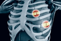
Using 3D-printed models based on chest CT scans allows clinicians to personalize surgical treatment for patients with multiple rib fractures -- reducing operating times and the risk of intraoperative complications, according to an article published online June 11 in the Journal of Cardiothoracic Surgery.
The researchers from Shijiazhuang Third Hospital in China developed a technique that relies on 3D printing technology to support presurgical planning for rib fracture repair. Prior research by a separate team of investigators from Taiwan demonstrated how 3D-printed models of rib structures helped boost the efficiency of simple rib fracture stabilization procedures. Rather than perform traditional rib stabilization procedures, which would have required large incisions in various locations, the group was able to perform minimally invasive surgery with the aid of 3D-printed models.
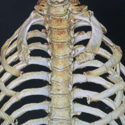
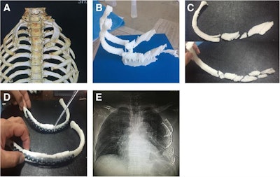 (A) Virtual 3D model of a patient's rib cage based on preoperative CT scans. (B) 3D-printed model of the fractured ribs. (C) 3D-printed model spliced and then reconstructed (D) onto titanium alloy rib-locking plates. (E) Postoperative chest x-ray showing restoration of rib structure. Images courtesy of Zhou et al. Licensed under CC BY 4.0.
In the current case report, first author Xue-Tao Zhou and colleagues expanded upon this earlier work and used 3D printing technology to facilitate presurgical preparation for several complex chest injuries involving multiple rib fractures. One case, for example, included a series of fractures along rib bones 2 through 11 caused by a motor vehicle accident.
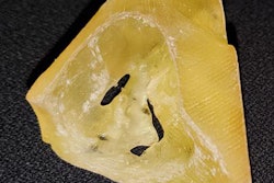
For all of the cases, the researchers acquired preoperative chest CT scans of the patients and then used computer software to make 3D reconstructions of the chest wall based on the scans. They subsequently input these reconstructions into 3D printing software and used a 3D printer to generate 3D-printed models of the fractured rib structures.
Having access to a model of the ribs allowed the group to determine preoperatively the precise size and shape of the titanium locking plates that the surgeons needed to attach onto the rib bones during surgery. Being able to individually tailor the titanium plates for each patient enhanced their fit onto the chest wall and improved the likelihood that the fractured ribs would heal properly, Zhou and colleagues noted.
"This method is more effective in terms of keeping the thoracic integrity and reducing the pulmonary complications. ... [It] is more beneficial for fitting the fixator perfectly at both ends of the rib fracture and leads to a more effective recovery of thoracic integrity," they wrote.
The preoperative application of 3D printing technology can enable clinicians to manually conform the titanium plates to rib structures before even commencing surgery, making it easier to complete the operation and minimizing the potential risk of injury, the authors continued.
"For some specific types of rib fractures, the preoperative application of 3D printing technology has potential significance in achieving precise and individualized treatment, but this method still needs more clinical experience to provide better services for patients," they concluded.


