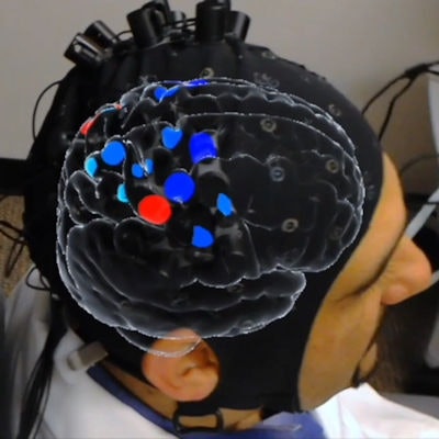
Researchers from Michigan have developed a new technique for real-time pain assessment on virtual 3D brain models using a combination of artificial intelligence (AI) algorithms and augmented reality (AR) technology. They detail their method in an article recently published in the Journal of Medical Internet Research.
Accurately gauging a patient's pain is often essential to proper diagnosis and treatment, yet standard pain assessment usually relies on questionnaires and scales that are subjective in nature and cannot be employed during procedures. Various groups have attempted to use neuroimaging such as functional MRI (fMRI) and PET to more objectively determine pain levels, but the sheer size and cost of conventional scanners make their regular clinical application impractical, first author Xiao-Su Hu, PhD, from the University of Michigan and colleagues noted.
In the feasibility study, Hu and colleagues tested a new approach to evaluating pain in a clinical setting. Their technique involved using functional near-infrared spectroscopy (fNIRS) to measure the varying concentrations of oxygenated and deoxygenated hemoglobin in the brains of 21 participants with orofacial pain (J Med Internet Res, June 2019, Vol. 21:6, e13594).
They applied several different AI algorithms to these data in order to decode the ongoing cascade of pain activity in the cortical region of the brain. The best of the algorithms -- an artificial neural network -- achieved an accuracy exceeding 80% for confirming the presence of pain. A different convolutional neural network logged the highest score for accurately predicting the site of pain at approximately 74%.
Seeking to apply this information in a clinical setting, the researchers developed proprietary software capable of generating a virtual 3D brain that displayed the recorded pain activity in different regions of the brain as colored flashes. They made the software compatible for use with an AR headset (HoloLens, Microsoft) so that they could visualize a personalized virtual brain pain map directly on the head of each participant.
The AR headset also displayed an animated human body alongside the virtual brain that indicated whether a participant felt pain on the right or left side of the body. Collectively, the AR presentation allowed clinicians to visualize the degree and general location of an individual's pain in real-time.
Virtual 3D model of the brain superimposed onto a person's head using AR technology. Colored areas within the brain model indicate varying degrees of oxygen saturation. Red areas on the animated human body beside the participant indicate regions of pain confirmed by AI. Video courtesy of Hu et al. Licensed under CC BY 4.0. Edited for length by AuntMinnie.com.
"In a true clinical environment, with such [a] framework, clinicians can better understand in an objective way [how] to determine when/where the patients are suffering from pain, especially when they cannot express [it] themselves. ... Such information will help clinicians decide when to intervene for addressing the pain or the immediate likelihood of it to occur," the authors wrote.
The technique may be viable, but it is still in the early stages of its implementation and will require further optimization before it can be used to assess pain conditions and neurologic disorders in the clinic, they concluded. The researchers remain hopeful that their technique "might turn into reality the goal of precisely 'seeing and believing' the biologic pain suffering of our patients in the doctor's office."


