Dear Advanced Visualization Insider,
Photon-counting CT (PCCT) continues to demonstrate its clinical utility across a broad range of clinical applications. For example, it could potentially be helpful as a new tool to opportunistically detect hepatic steatosis -- a condition linked to significant health complications such as liver fibrosis and cirrhosis, metabolic syndrome, diabetes, and coronary artery disease.
Researchers led by Dr. Fides Schwartz of Duke University have found that the technology performs just as well as MRI for identifying hepatic steatosis. You can get all of the details by viewing this edition's Insider Exclusive.
Speaking of PCCT, the technology was also recently shown to enable 25% lower contrast volume for coronary CT angiography (CCTA) exams without impacting image quality,
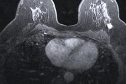
In other advanced visualization news, a machine-learning algorithm that analyzes radiomics features on dynamic contrast-enhanced MRI demonstrated potential for characterizing breast lesions. Also, an MRI radiomics-based nomogram offers promise for predicting treatment response in patients with colorectal cancer.
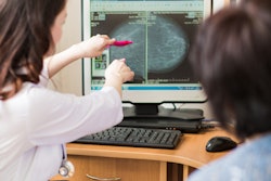
What's more, a nomogram based on radiomics and other ultrasound features on automated breast volume scanning can enable noninvasive and accurate analysis of axillary lymph node status in women with early invasive breast cancer. Making use of CT radiomics, another artificial intelligence algorithm could assess the risk of coronary artery disease in diabetic patients.
In 3D printing developments, models of retroperitoneal tumors were deemed effective for planning surgery for children and to communicate about the procedure to their parents. Meanwhile, a genitourinary 3D modeling and printing process based on MRI exams can help in planning pediatric urologic surgeries -- and potentially even other types of procedures.
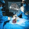
A team from Japan also shared their success with a novel approach to ultrasound-guided fine-needle biopsy for solid pancreatic tumors. In addition, CT-guided percutaneous biopsy was judged to be effective for obtaining tissue samples for soft tissue and bone mass biopsies. Furthermore, MRI-guided stereotactic body radiotherapy is better than CT guidance for reducing toxic effects of the treatment, according to a recent study.
Incorporating bone-suppression functions can improve detection of subtle pulmonary lesions on temporal subtraction digital chest radiographs. A research group has also developed a 3D imaging prototype that can visualize real-time radiation dose in patients undergoing radiation therapy.
Is there a story you'd like to see covered in the Advanced Visualization Community? Please feel free to drop me a line.


