Dear AuntMinnie Member,
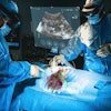
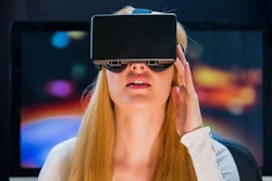
Can virtual reality (VR) help improve patient experience during interventional radiology procedures? Yes, it can, according to a new study that we’re featuring in this Advanced Visualization Insider.
Researchers from Temple University implemented a VR-based approach that provided patients with a simulated tropical island environment during their peripherally inserted central catheter procedure or fine-needle aspiration thyroid biopsy. They found that VR reduced both patient pain and anxiety – without increasing procedure time. Click here for our report.
But augmented reality (AR) may also have a role in interventional radiology. A group at McMaster University in Ontario, Canada, reported that AR could improve the accuracy of needle placements during image-guided tumor ablation procedures.
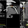
A study presented at ECR 2024 in Vienna described how a deep-learning algorithm can quantify patients’ subcutaneous fat tissue levels on lung cancer screening CT exams. These results could help clinicians tailor patient care, according to the group from Germany.
In other advanced visualization developments, a research team has developed a deep-learning algorithm to automatically segment neonatal brains on MR images. Their model performed well on both high- and low-resolution MRIs. Get all of the details here.
The adoption of AI-based radiomics analysis has been held back in part by concerns over the reproducibility of results. Standardized image processing techniques developed by a multinational team may be able to help, however. After applying eight software “filters” on a multimodal set of imaging datasets, the researchers concluded that 94% of radiomics features were reproducible. Click here for our coverage.
Is there an advanced visualization story you'd like to see covered? Please feel free to drop me a line.


