Dear Advanced Visualization Insider,
A new 3D intraoperative imaging technology developed at the University of Toronto is showing promise for providing surgeons with submillimeter spatial resolution and soft-tissue visibility in real-time.
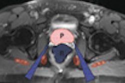
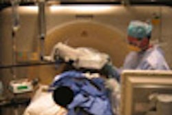
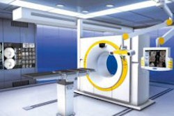
The technology, shown at the recent American Association of Physicists in Medicine (AAPM) meeting in Minneapolis, combines a conebeam CT system and a mobile C-arm. It can acquire a full volume image in a single rotation and is currently in trials with patients undergoing brachytherapy, as well as chest, breast, spine, and head and neck surgery, according to presenter Jeffrey Siewerdsen, Ph.D.
Our coverage of the new 3D intraoperative technology is the subject of this month's Insider Exclusive article. You have access to the story before it is published for the rest of our AuntMinnie.com members. To learn more, click here.
In other articles we're featuring this month in the Advanced Visualization Digital Community, radiologists failed to better the performance of computer algorithms in measuring lung nodules, according to a presentation at Stanford University's 2007 International Symposium on Multidetector-Row CT in San Francisco. For coverage of that presentation, click here.
In other coverage from that conference, we report on a talk by Stanford's Laura Pierce, who shares strategies on retaining those ever-valuable 3D technologists. For that article, click here.


