Sunday, November 27 | 11:15 a.m.-11:25 a.m. | SSA15-04 | Room N229
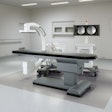
Researchers from NTT Medical Center in Tokyo have developed a new compact intraoperative MRI system to provide high-quality images in the surgical suite.Dr. Akio Morita, PhD, and colleagues have been working on the compact MRI system since October 2007. Completing the system took about three years, and it was installed at the medical center in September 2010, Morita said. A clinical trial is under way to evaluate the technology, which has been used in approximately 10 cases so far.
The compact MRI system features a 0.23-tesla magnet and gantry that weighs a total of 2.8 tons. To obtain high-quality images in the neurosurgical setting, the researchers also developed a new field-of-view head coil to provide a uniform magnetic field for the entire cranium. The coil can be attached to the head clamp system during surgery.
Morita and colleagues also developed a new canopy-style radiofrequency shield system to minimize noise and maintain a high signal-to-noise ratio in the operating suite.
The average image acquisition time has been approximately 52 minutes, and so far there have been no clinical complications or system failures, Morita said.
"Currently, we are applying this system to cranial-brain surgery, especially for brain tumors such as glioma, pituitary adenoma, metastasis, and so on," Morita added. "We have not applied this system for cerebrovascular or craniofacial surgery yet, but we are planning to do so."
The system can be used for head and neck and upper C-spine surgery as it is currently configured, but those applications have not yet been tried, Morita said.
The device and complementary coil systems are also under further development for orthopedic limb and breast cancer surgery. "Spine surgery is also in our scope," Morita added.
The compact MRI system is built and distributed by Yoshida Manufacturing.


