Sunday, November 26 | 1:00 p.m.-1:30 p.m. | IN205-SD-SUB3 | Lakeside, IN Community, Station 3
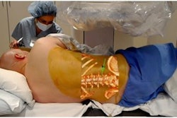
Radiologists can design and develop realistic simulation phantoms for fluoroscopic-guided shoulder arthrography by combining 3D printing with molding techniques, according to U.S. researchers.Image-guided procedures call for an extensive understanding of 3D anatomic relationships, involve potential complications, and often require the use of radiation. And yet there are few realistic ways to cultivate procedural skills apart from real-life experiences.
"I felt the need for a process that could bridge the gap between the theoretical understanding of how a procedure is done and [the act of] actually performing the procedure on a patient," Dr. Ramin Javan told AuntMinnie.com.
In this poster presentation, Javan and colleagues from George Washington University Hospital in Washington, DC, will detail how they created a 3D model containing a joint space that radiologists could use to safely practice fluoroscopy-guided shoulder arthrography.
The researchers will explain exactly how to design and develop 3D-printed bone structures and joint capsules, which entails a complicated process. They will also offer solutions to several challenges that arise when producing 3D-printed models, especially when constructing models for presurgical planning.
"3D prints can be customized based on the designer's educational goals and even be patient-specific," Javan said. "This [practice] can improve the confidence of trainees, increase the chance of success, decrease procedure time and hence radiation dose, decrease patient discomfort, and possibly decrease complication rates."
