Thursday, November 29 | 11:50 a.m.-12:00 p.m. | SSQ17-09 | Room N229
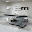
Mobile CT scanning combined with an artifact-reduction algorithm may enhance the visualization of soft tissue for radiotherapy, according to this Thursday session.The researchers, led by Jeff Siewerdsen, PhD, from Johns Hopkins University, tested the viability of a relatively new mobile CT scanner for image-guided brachytherapy -- a form of radiotherapy often used to treat cervical and breast cancer. Applying the technique to realistic phantoms of the head, chest, abdomen, and pelvis, the group found that the machine provided sufficiently high-quality 3D imaging for tracking needles and applicators within soft tissue.
Presenter and doctoral student Nicole Chernavsky, Siewerdsen, and colleagues also identified two main forms of image artifacts that diminished soft-tissue visibility: stitching artifacts seen on axial views and windmill artifacts associated with high-frequency objects such as bone or metal instruments. Adding a metal artifact reduction algorithm helped minimize the appearance of these artifacts.
New mobile CT scanners could help improve the precision and safety of CT-guided interventions, including brachytherapy and image-guided spinal surgery, Siewerdsen told AuntMinnie.com.
"Overall, the imaging performance [of the mobile CT scanner] was suitable to interventional guidance for a variety of bone and soft-tissue interventions, and the study helped to identify important areas of improvement in technique protocol, reconstruction method, and artifact reduction," Chernavsky said.


