Dear AuntMinnie Member,
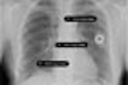
One of the next frontiers for computer-aided detection (CAD) technology is the evaluation of pulmonary embolism (PE) in CT studies. Imaging professionals are hoping that CAD can provide many of the benefits that it is already delivering in mammography screening applications.
But recent studies indicate that while CAD has potential, it is also being plagued by some of the same challenges that breast imagers are experiencing. So reports staff writer Erik L. Ridley for our Advanced Visualization Digital Community.
In one study, Canadian researchers found the CAD software they tested to be easy to use, but it produced too many false-negative results. They recommended improvements to the technology before they could recommend its routine use.
U.S. researchers, however, achieved better results in their use of CAD to detect and assess PE and predict embolism severity. While the group had to cope with a number of false positives, they found that the CAD algorithm produced good agreement with measures of pulmonary obstruction as produced by radiologists.
Another U.S. group likewise reported very strong results for CAD in detecting acute pulmonary emboli in patients without significant pulmonary disease.
Finally, German researchers also enjoyed good results for CAD in their study of more than 130 patients. While the group also reported a large number of false positives, they did not experience any false negatives in patients with PE.
Get the details on all four studies by clicking here, or visit our Advanced Visualization Digital Community at av.auntminnie.com.

