Tuesday, December 3 | 11:40 a.m.-11:50 a.m. | SSG06-08 | Room S406A
Artificial intelligence (AI) has the potential to produce standardized, accurate x-ray reports in a timely manner that are easy to comprehend in the clinical setting.In this presentation, Dr. Vasantha Kumar Venugopal, a radiologist at the Institute of Brain and Spine in New Delhi, will show how he and fellow researchers evaluated the proficiency of AI-generated chest x-rays compared to radiologist-generated clinical reports.
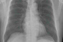
Approximately 300 chest x-rays were performed on a conventional x-ray system retrofitted with digital radiography. The anonymized chest x-ray images were analyzed by a deep learning-based chest x-ray algorithm designed to detect abnormalities and automatically generate clinical reports.
The algorithm features a combination of multiple classification, detection, and segmentation neural networks designed to identify 75 different radiological findings and determine the location of the abnormalities. The networks themselves were trained using approximately 1 million chest x-rays from multiple data sources.
In the study, the algorithm-generated reports were deemed as accurate as the radiologists' reports in 79% of cases. In 5% of the cases, the algorithm produced reports that were more accurate or more clinically appropriate than those of the radiologists.
Conversely, the algorithm-created reports had significant diagnostic errors in 6% of cases, while in 9%, the algorithm created reports that were found to be clinically inappropriate or insufficient, even though the significant findings were correctly identified and localized.
The reports automatically generated by the algorithm resulted in "good comparability" with "high accuracy, paving the way for a new potential deployment strategy of AI in radiology," the group concluded.

