Wednesday, November 30 | 10:30 a.m.-10:40 a.m. | SSK17-01 | Room S404AB
In this scientific session, researchers will reveal how an automated method based on deep learning shows promise for segmenting and measuring the volume of lymph node clusters.The presence of enlarged lymph nodes signals the onset or progression of a malignant disease or infection, and lymph node segmentation is an important yet challenging problem in medical image analysis, according to senior author Dr. Ronald Summers, PhD, of the U.S. National Institutes of Health (NIH) Clinical Center.
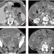
In the thoracoabdominal body region, neighboring enlarged lymph nodes often spatially collapse into "swollen" lymph node clusters. However, accurate segmentation of these lymph node clusters is complicated by the noticeably poor intensity and texture contrast among neighboring lymph nodes and surrounding tissues, he said.
"Furthermore, the boundaries between distinct agglomerated lymph nodes are highly ambiguous," Summers told AuntMinnie.com. As a result, lymph node diameter measurement in the thoracoabdominal region is subject to human error and high interobserver variability.
To help, the NIH team developed a fully automated method for thoracoabdominal lymph node cluster segmentation that integrates deep learning technology with graph-based structured optimization techniques. In testing, the method yielded "state-of-the-art" segmentation and volume measurements results, according to the group, which included presenter Isabella Nogues and Le Lu, PhD.
"Such results are promising for the development of [lymph node] imaging biomarkers based on volumetric measurements, which may lay the groundwork for improved [Response Evaluation Criteria in Solid Tumors (RECIST)] lymph node measurements," Summers said.
Stop by this Wednesday morning talk to learn more.


