The PET agent 2-(fluorine-18) fluo-2-deoxy-D-glucose (18-FDG) has been the most studied
for evaluation of bronchogenic carcinoma. 18-FDG competes with glucose for facilitated
transport into tumor cells and also competes with glucose for phosphorylation by
hexokinase. Unlike glucose, however, the phosphorylated form is not further metabolized,
it becomes trapped within the cell with little back diffusion or degradation. Uptake of
FDG therefore reflects regional rates of exogenous glucose utilization. The agent is useful for imaging bronchogenic carcinoma because lung
tumor cells have an increased cellular uptake of glucose due to an increased number of
surface transport proteins and a higher rate of glycolysis in comparison to non-neoplastic
cells (ie: increased metabolic rate in lung cancer cells).
FDG PET imaging can have a significant impact on patient
management by heightening suspicion for pulmonary malignancy, identifying unsuspected
sites of disease, and by guiding selection of a biopsy site. Similarly, a negative PET
scan indicates a low likelihood for malignancy and supports the use of conservative
management and follow-up [40]. In a study evaluating the impact of FDG PET exam results in
the clinical management of patients with suspected or known thoracic malignancies PET
scans influenced treatment in 65% of patients and offered new information in 85% [40].
FDG PET imaging can be used to evaluate indeterminate solitary pulmonary nodules to
determine whether the nodule is benign or malignant. A standardized uptake ratio (SUR) is
used to determine if a lesion has increased 18-FDG activity. The SUR normalizes the amount
of FDG accumulation in a region of interest (ROI) to the total injected dose and the
patient's body weight. It provides a means of comparison of FDG uptake between patients
[41]. The SUR is calculated by dividing the mean activity within a selected region of
interest (in mCi/ml) by the injected dose (in mCi/kg). Modifications of the SUR that may
improve the semiquantitative evaluation of FDG uptake include using body surface area or
the lean body weight instead of the weight of the patient; this is significant because the
distribution of FDG is higher in muscle than in fat [44]. It is also important to remember
that SUR values may change with time after FDG injection; thus, the time of acquisition
after FDG injection must be standardized for the values to be useful [25]. Other factors
which can affect the SUV include partial-volume effects and the blood glucose level at the
time of injection [46].
SUR= Mean selected region activity (mCi/ml)/injected dose (mCi)/body weight (kg)
An SUR greater than 2.5 has been shown to be very sensitive and specific for malignant
lesions [13]. Visual analysis of the amount of uptake within a lesion has also been shown
to be effective in differentiating benign from malignant lesions [13,47]. Uptake greater
than blood pool activity indicates a malignant lesion, while activity equal to or less
than mediastinal blood pool suggests a benign lesion [47]. In fact, visual analysis may be
more sensitive for nodules smaller than 1.5 cm in size (although, the improved sensitivity
comes at a decreased specificity) [47].
High sensitivities (82-100%), specificities (67-100%), and accuracy (79-94%) have been
reported for the identification of pulmonary malignancy using FDG PET imaging [25,47,50,52].
However, the high incidence of cancer in the many of the study groups may
contribute to the high reported
sensitivities. In a prospective multicenter study for the evaluation of pulmonary nodules
(larger than 7 mm) felt to be indeterminate for malignancy based upon their conventional
imaging appearance, FDG PET had a sensitivity of 92% and a specificity of 90% using SUR
data [47]. Visual analysis of the images demonstrated a sensitivity of 98%, but a lower
specificity of 69% [47].
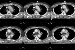
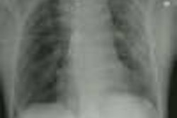
Example: The CT scan
in this patient demonstrated a small nodule in the left upper lobe (black
arrow). PET-FDG images demonstrate
very intense accumulation of the tracer within the lesion (white arrow),
which was a non-small cell lung cancer. (Case courtesy of H. Page McAdams
MD, Department of Radiology, Duke University Medical Center)
False positive results are not uncommon and can be seen with
infectious granulomas (tuberculosis, histoplasmosis, aspergillosis, cryptococcus, and
inflammatory pseudotumor) [36,50] or inflammatory processes (sarcoid, Wegener's),
rheumatoid nodules [13,15], and an aggressive neurofibroma. [8]
Although uncommon, false negative exams can occur in three settings
[41]:
1- A neoplasm with low metabolic activity: Bronchoalveolar cell carcinoma may have a
lower uptake value and can cause a false-negative result in up to 57% of cases
[18,21,47,50]. Another pulmonary neoplasm which frequently has a negative FDG PET exam
(SUR < 2.5) is a carcinoid tumor (up to 85% of cases) [42,43,50]. Although less common,
other NSCLC's such as squamous cell carcinoma and adenocarcinoma may also fail to
demonstrate significant FDG accumulation [38,47]
2- Small lesions (under 1 cm to 1.5 cm in size) or lesions with a physically small
histologic complement of malignant cells: Lesions as small as 1 cm can be accurately
evaluated, but the evaluation of smaller lesions may result in false negative results due
to spatial resolution limitations of PET imaging (approximately 5 mm) and respiratory
motion [13,21]. FDG PET has shown a decreased sensitivity (80%) for malignancy for lesions
smaller than 1.5 cm [41,47]. The ability to characterize smaller lesions is in part
dependent on the degree of tracer accumulation within the lesion. Even very small lesions
can be correctly characterized if they accumulate enough FDG to become visible.
Sensitivity for lesions between 5-8 mm in size is about 50% [41].
3- Hyperglycemia [47]: Competitive inhibition from high serum glucose levels appears to
hinder FDG uptake in some cases. This effect is most important with acute hyperglycemia
while a chronically raised glucose level inhibits tumor uptake minimally (about 10%) [41].
Based upon the present data available- a lesion that is hot on PET should be considered
malignant until proven otherwise. A lesion that is PET negative has a low probability for
being a malignancy (under 5% [37]). However, one must consider all characteristics of a
lesion prior to discounting its malignant potential. Follow-up exams should be performed
on PET negative nodules to ensure stability [37]. If the lesion grows, further evaluation
with tissue diagnosis should be obtained [37].
Nuclear medicine gamma cameras equipped with ultrahigh energy collimators have also
been used to perform SPECT images of 18-FDG in patients with suspected lung cancer.
Overall sensitivity of SPECT FDG is 77% to 81% for identifying malignant pulmonary
lesions (sensitivity has been report to be 100% for lesions larger than 2 cm [24]).
Specificity is 91-100%. Small malignancies (less than 2 cm in size) are not reliably
characterized with this imaging technique (sensitivity for lesions less than 2 cm is only
20-50%). The small number of patients studied and the
high incidence of malignancy in the study groups (60-75%) make any conclusions about the
true sensitivity of SPECT 18-FDG premature. [10]
Nuclear medicine dual detector gamma cameras have also been modified to permit
coincidence detection. In general, these cameras have a limited intrinsic detector
efficiency for the high energy FDG emissions which results in poor counting statistics.
Non-true coincidence detection also contributes to image noise. None-the-less, pulmonary
lesions larger than 1 cm seem to be well detected due to the relatively low background
activity of the lungs. Lesions within the deep mediastinum and abdomen less than 1.5 cm in
size, are less well identified [19].
An accurate assessment off the efficacy of chemotherapy and radiation therapy might
prove of enormous value in directing therapy for patients with advanced stage NSCLC [45].
Normalization of FDG uptake after treatment indicates a good response to therapy [45].
However, it may be important to differentiate decreased FDG uptake from absence of uptake
[45]. Decreased uptake may indicate only a partial response to treatment resulting from
destruction of sensitive cells, while resistant cells continue to be metabolically active
[45].
PET FDG imaging may also provide prognostic information regarding patient outcome [29].
Preliminary data indicate that patients with positive PET scans following initial lung
cancer therapy have significantly worse progoses than patients with negative results [29].
Unfortunately, a decrease in FDG uptake may only indicate a partial response to therapy,
while normalization after treatment appears to be a good prognostic indicator [41]. In one
study, all patients with negative PET exams following treatment were alive at two years
(even in the presence of non-specific radiologic changes), while 50% of patients with
residual hypermetabolism had died during the same two year period [41,45]. Further
evaluation will be required to determine if changes in therapeutic regimens should be made
based upon positive PET FDG results after first-line treatment.
Radiation therapy can result in inflammatory changes that may be difficult to
differentiate from recurrent tumor. Generally, radiation produces a diffuse, mildly
elevated FDG accumulation within the tissues included in the radiation port [41]. This
activity should decrease over time and scans will be most reliable when a year or more has
passed from the last radiaiton treatment [41]. A SUR of 2.5 still appears to be accurate
in differentiating benign changes from tumor [41].
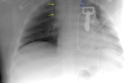
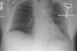
Example: PET imaging has also been used to document response to
treatment. In the case below, intense tumor (black arrow) and nodal (blue arrow) uptake
can be seen to decrease following chemotherapy. (Case courtsey of Dr. David Mankoff,
Division of Nuclear Medicine, University of Washington, Seattle)
A major problem in the follow-up evaluation of patients treated for lung cancer is
distinguishing post-treatment changes from recurrent tumor [48]. Presently, tumor
recurrence is often not diagnosed until the disease has progressed to the point of marked
enlargement of questionable abnormalities [41]. PET FDG imaging is very sensitive and
highly accurate in distinguishing recurrent malignancy from scarring/fibrosis
[13,41,48,49]. FDG PET imaging has been shown to have a sensitivity 98-100% for the
differentiation of post-treatment change from tumor recurrence [48]. In a prospective
evaluation of patients treated for NSCLC [48], FDG PET was able to correctly identify
recurrent disease in all patients (sensitivity 100%) [48]. In this same group, CT imaging
was non-contributory in 28% of cases at the time of FDG PET diagnosis [48]. False positive
exams can occur in association with infection and radiation pneumonitis [48]. Positive FDG
PET exams due to radiation pneumonitis can be seen in up to 4% of patients receiving XRT
therapy. The duration of abnormality associated with radiation pneumonitis is variable and
exams can remain positive (SUV greater than 2.5) for 6 to 15 months [48]. FDG PET findings
associated with radiation pneumonitis should show decreased activity over time [48]. It is
probably best not to perform FDG PET imaging earlier than 6 months after XRT in order to
reduce the likelihood of false-positive PET results [48].
FDG-PET can also assist in identifying hypermetabolic foci within radiographic
abnormalities to better direct tissue biopsy [45].
Screening for recurrence is another potential use of FDG PET imaging [48]. In a group
of patients with NSCLC treated for cure, FDG PET screening was able to document the
presence of recurrent tumor in all patients, whereas the finding was missed by CT in 44%
[48]. If detected sooner, retreatment could be instituted in an effort to obtain prolonged
disease control [48]. Further studies will be required to determine if improved detection
of recurrent on FDG PET screening can result in improved patient survival [48].
(39) AJR 2000; Boiselle PM, et al. Lung cancer detection in the 21st century:
Potential contributions and challenges of emerging technologies. 175: 1215-1221
(No abstract available)
(40) Chest 2000; McCain TW, et al. The usefulness of
positron emission tomography in evaluating patients for pulmonary malignancies.
118: 1610-1615
(46) J Nucl Med 2001; Avril N, et al. Glucose
metabolism of breast cancer assessed by 18F-FDG PET: Histologic and
immunohistochemical tissue analysis. 42: 9-16
(48) Eur Respir J 1999; Bury T, et al. Value of FDG-PET
in detecting residual or recurrent nonsmall cell lung cancer. 14: 1376-1380
(49) J Clin Oncol 2001; Kalff V, et al. Clinical
impact of 18F fluorodeoxyglucose positron emission tomography in patients with
non-small-cell lung cancer: A prospective study. 19: 111-118
(50) Chest 2001; Dunagan DP, et al. Staging by
positron emission tomography predicts survival in patients with non-small cell
lung cancer. 119: 333-339
(51) Chest 2001; Hauber HP, et al. Positron emission
tomography in the staging of small-cell lung cancer. A preliminary study. 119:
950-954
(52) J Cancer Res Clin Oncol 2000; Ilknur AK, et al.
Positron emission tomography with 2-[18F] fluoro-2-deoxy-D-glucose in oncology:
Part II: The clinical value in detecting and staging primary tumours. 126:
560-574