Pulmonary embolsim in a patient with left external iliac vein thrombosis
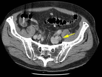
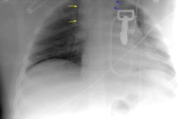
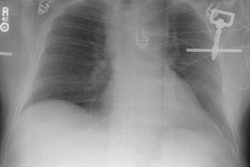
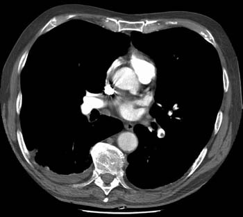
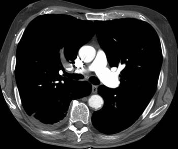
The patient shown below was post op from repair of an abdominal aortic aneurysm. The CT PE exam demonstrated emboli to the left lower lobe and right upper lobe (large clot) pulmonary arteries. The DVT portion of the exam detected clot in the left iliac vein- a finding which would not have been evident on lower extremity ultrasound. In patients that require an inferior vena caval filter the identification of clot within the iliac vessels or IVC can aid in the angiographic approach to filter placement.
Clot is evident as a filling defect in the left lower lobe pulmonary artery.
Clot completely occludes the right upper lobe pulmonary artery
A left external iliac vein clot was identified during the CT DVT exam (yellow arrow)
