Acute pulmonary embolism: role of helical CT in 164 patients with intermediate probability at ventilation-perfusion scintigraphy and normal results at duplex US of the legs.
Ferretti GR, Bosson JL, Buffaz PD, Ayanian D, Pison C, Blanc F, Carpentier F, Carpentier P, Coulomb M
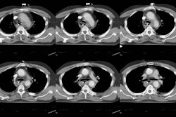
PURPOSE: To assess prospectively the clinical effectiveness of helical computed tomography (CT) in the evaluation of patients with unresolved suspicion for pulmonary embolism (PE). MATERIALS AND METHODS: Helical CT was performed in 164 consecutive patients suspected of having acute PE, intermediate probability at ventilation-perfusion (V-P) scintigraphy, and normal findings at duplex ultrasonography (US) of the legs. Fifteen patients also underwent pulmonary angiography. Helical CT results were analyzed immediately to help plan anticoagulant treatment. If helical CT did not show PE, anticoagulant treatment was not indicated. Clinical outcome for these patients was assessed during 3-month follow-up. RESULTS: In 40 (24.4%) of 164 patients, the diagnosis of PE was based on results at helical CT (n = 39) or pulmonary angiography (n = 1). Repeated Doppler US of the legs depicted one thrombus in the calf of three patients with normal results at helical CT that could have been responsible for PE. During 3-month follow-up, three patients experienced recurrent PE (one death, two recurrences). Therefore, PE occurred in six (5.4% [95% confidence interval, 1.3%, 9.7%]) of 112 patients with normal findings at helical CT who did not receive anticoagulant treatment. CONCLUSION: Findings at helical CT allowed accurate diagnosis of acute PE in patients with intermediate probability at V-P scintigraphy and without deep venous thrombosis at duplex sonography of the legs.


