Spiral CT appearance of resolving clots at 6 week follow-up after acute pulmonary embolism.
Van Rossum AB, Pattynama PM, Tjin A Ton E, Kieft GJ
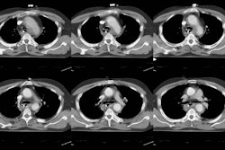
PURPOSE: The purpose of this work is to describe the spiral CT appearance of resolving clots at 6 week follow-up in patients with acute pulmonary embolism (PE). METHOD: Nineteen patients with acute PE initially identified with spiral CT scan underwent repeat CT examinations at 6 week follow-up after the start of anticoagulant therapy. The appearances of the clots on the initial CT scan and follow-up CT scan were analyzed. RESULTS: Normalization of the pulmonary arteries at follow-up was seen in six patients (32%) only. Residual abnormalities were present in 13 of 19 patients (68%). Resolving clots were seen as eccentric wall-adherent filling defects (22%) or filling defects with central contrast material (3%). CONCLUSION: Resolving clots after acute PE can be seen with follow-up CT scan in the majority of patients. It is important to be familiar with these findings.


