Parenchymal and pleural findings in patients with and patients without acute pulmonary embolism detected at spiral CT.
Shah AA, Davis SD, Gamsu G, Intriere L
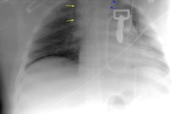
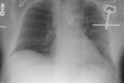
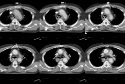
PURPOSE: To compare the frequencies of parenchymal abnormalities and pleural effusions in patients with and patients without acute pulmonary embolism (PE) detected at spiral computed tomography (CT). MATERIALS AND METHODS: Contrast material-enhanced spiral CT scans obtained in 92 patients clinically suspected of having acute PE were retrospectively reviewed. The presence or absence of parenchymal abnormalities and pleural effusions was noted. The presence of filling defects consistent with central or peripheral PE was recorded. RESULTS: Twenty-eight patients had CT evidence of PE. Central emboli were evident in 27 (96%) of these patients; 23 (82%) had concomitant central and peripheral emboli, and four (14%) had only central emboli. One patient had an isolated subsegmental clot. Parenchymal abnormalities were seen in 24 (86%) patients with PE and 56 (88%) patients without PE. Atelectasis, the most common finding, was present in 20 (71%) patients with PE and 41 (64%) patients without PE. The only parenchymal abnormality significantly associated with PE was peripheral wedge-shaped opacity, which was seen in seven (25%) patients with PE and three (5%) patients without PE (odds ratio, 6.78; 95% CI = 1.60, 28.62). Pleural effusions were seen in 16 (57%) patients with PE and 36 (56%) patients without PE. In 25 (39%) patients without PE, there were additional CT findings that might suggest an alternative explanation for the acute clinical presentation. CONCLUSION: Parenchymal and pleural findings at CT are of limited value for differentiating patients with PE from those without PE.
PMID: 10189464, UI: 99205462


