Traumatic aortic injuries in children: radiologic evaluation.
Lowe LH, Bulas DI, Eichelberger MD, Martin GR
OBJECTIVE: The purpose of this study was to evaluate the radiographic
findings in children with traumatic aortic injuries and discuss the imaging
techniques currently available for diagnosis. MATERIALS AND METHODS: A
retrospective review of 10,886 children examined because of blunt trauma
from 1987 to April 1996 identified seven patients (0.064%) who sustained
traumatic aortic injuries. The mechanism of injury, location of aortic
injury, additional injuries suffered, trauma scores, sequences of radiologic
evaluation, imaging findings, treatment, and outcome were recorded for
each child. RESULTS: Six children had pathologically proven aortic ruptures,
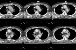
and the remaining child had an intimal injury diagnosed with contrast-enhanced
helical CT and confirmed with transesophageal echocardiography. All seven
children were victims of motor vehicle accidents (six passengers, one pedestrian),
all had injuries of the aortic isthmus, and all had additional severe injuries.
The mean trauma score, injury severity score, and probability of survival
were 14, 39, and 75%, respectively. Imaging techniques included chest radiography
(n = 7), conventional CT (n = 1), helical CT (n = 3), aortography (n =
2), and transesophageal echocardiography (n = 3). The initial outcomes
included death (n = 1), paraplegia (n = 1), paraparesis (n = 2), and recovery
without morbidity (n = 3). CONCLUSION: Traumatic aortic injuries are rare
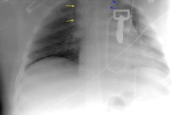
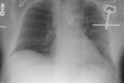
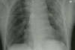
in children. The most common findings on plain films are a left apical
cap, pulmonary contusion, aortic obscuration, and mediastinal widening.
Helical CT and transesophageal echocardiography can be used in the diagnosis
of traumatic aortic injuries in children.


