CHICAGO -- MRI has shown that skeletal muscle loss (i.e., sarcopenia) is an early risk factor for cognitive decline in older adults, according to research presented December 3 at the RSNA meeting.
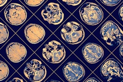
The findings suggest that brain MR imaging that quantifies sarcopenia via an assessment of the temporalis muscle could serve as a way to predict future incidence of dementia, according to a group led by Kamyar Moradi, MD, of Johns Hopkins Medicine in Baltimore, MD.
 Kamyar Moradi, MD.
"Measuring temporalis muscle size as a potential indicator for generalized skeletal muscle status offers an opportunity for skeletal muscle quantification without additional cost or burden in older adults who already have brain MRIs for any neurological condition, such as mild dementia," Moradi said in an RSNA statement.
Skeletal muscle mass begins to decrease as people age and is often seen in those with Alzheimer's disease, the team noted. Previous research has shown that temporalis muscle thickness and area can indicate muscle loss throughout the body; the muscle is located in the head and moves the lower jaw.
Moradi and colleagues conducted a collaborative study between the radiology and neurology departments at Johns Hopkins that included data from 621 cognitively healthy participants taken from the Alzheimer’s Disease Neuroimaging Initiative. They manually segmented images of the temporalis muscles and calculated the sum cross-sectional area of these muscles; they then categorized study participants into a large cross-sectional area group and a small cross-sectional area group and assessed dementia incidence, change in cognitive and functional scores, and brain volumes over a median follow-up period of 5.8 years.
Overall, the team found that a smaller temporalis muscle was associated with a higher risk of Alzheimer's disease dementia (hazard ratio, 1.59, with 1 as reference). Smaller temporalis muscles were also linked to a greater decrease in memory composite score, functional activity questionnaire score, and structural brain volumes, it noted.
"We found that older adults with smaller skeletal muscles are about 60% more likely to develop dementia when adjusted for other known risk factors," said the study's co-senior author Marilyn Albert, PhD, in the statement.
The study results indicate that using MRI to assess the temporalis muscle could be an effective way to track muscle loss as people age, allowing for interventions such as physical activity, resistance training, and nutrition, the group concluded.
