Rini JN, Leonidas JC, Tomas MB, Palestro CJ.
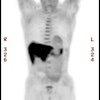
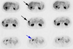
The purpose of this study was to compare (18)F-FDG PET to CT for evaluating the spleen during the initial staging of lymphoma. METHODS: Seven patients with newly diagnosed lymphoma underwent (18)F-FDG PET and CT. Splenic uptake of (18)F-FDG, diffuse or focal, greater than hepatic uptake was interpreted as consistent with tumor. CT demonstrating a positive splenic index or focal hypodensities was classified as positive for tumor. PET and CT results were compared with final diagnoses, which were confirmed surgically for 6 patients and at autopsy for 1 patient. RESULTS: Five of 7 patients had lymphomatous involvement of the spleen. (18)F-FDG PET was true-positive for all 5 patients with splenic disease and true-negative for both patients without splenic disease. CT, in contrast, was true-positive for 4 of the 5 patients with splenic disease and false-positive for the 2 patients without splenic disease. The accuracies of (18)F-FDG PET and CT for evaluating the spleen were 100% and 57%, respectively. CONCLUSION: (18)F-FDG PET correctly identified all patients with and without splenic disease and was superior to CT for this purpose.
