Dear AuntMinnie Member,
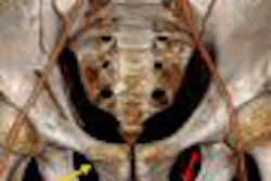
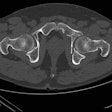
Acetabular fractures of the pelvis are relatively rare, but when they do occur they can produce serious complications such as blood clots, nerve injuries, and infections. Fortunately, radiologists are discovering that advanced visualization techniques -- specifically 3D volume-rendered CT -- offer a less invasive approach to assessing acetabular fractures than previous techniques.
That's according to an article we're featuring this week in our Musculoskeletal Imaging Digital Community by staff writer Shalmali Pal. The story details a presentation at last month's RSNA meeting in which U.S. researchers discussed their use of 3D volume-rendered CT for acetabular assessment.
The researchers found that using the 3D CT technique enabled them to eliminate additional imaging tests, saving patients both time and radiation dose. Learn more about how they did it by clicking here.
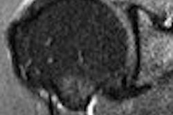
In another article in the community, New Zealand researchers have used MRI to identify high-grade bone edema in patients with rheumatoid arthritis (RA) who are undergoing orthopedic surgery. MRI can demonstrate signs of disease with the potential to erode bone, giving clinicians an early warning that aggressive treatment is needed. Get more details by clicking here, or visit our Musculoskeletal Imaging Digital Community at msk.auntminnie.com.
In other news, the recent shortage of technetium-99m may be coming to an early resolution. The Canadian parliament this week passed a resolution that would temporarily clear up the regulatory snafu that forced operators of a Canadian nuclear reactor to shut down -- halting the supply of molybdenum-99, the raw material for technetium. Get the latest on the story by clicking here.

