Physicians who perform percutaneous vertebroplasty should keep a watchful eye on treated vertebrae that exhibit a cleft pattern as these cases may be prone to new compression fractures, according to a paper in the December American Journal of Roentgenology.
Dr. Noboru Tanigawa and colleagues from the department of radiology at Kansai Medical University in Osaka, Japan, hypothesized that different cement patterns in treated vertebrae have an impact on the frequency of new compression fractures. They tested this theory in 76 patients with 266 osteoporosis-related fractures who underwent vertebroplasty.
The treatment protocol included presurgical x-rays, vertebral MR images, and CT scans. Vertebroplasty was done under CT and fluoroscopic guidance (Advantx LCA plus ACT, GE Healthcare, Chalfont St. Giles, U.K.). Twenty grams of methyl methacrylate (PMMA) powder, blended with 10 mL of liquid methyl methacrylate to a "toothpaste-like consistency," was injected into the vertebral body until adequately filled (AJR, December 2007, Vol. 189:6, pp. W1-W5).
A visual analogue scale (VAS) from 0 to 10 (worst pain) was used to assess the severity of postprocedural pain. Follow-up was done on an inpatient and outpatient basis with results based on a median of 16.7 months.
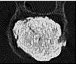
Vertebral bodies were classified as either cleft type (vertebrae with compact and solid cement filling) or trabecular type (vertebrae with sponge-like cement filling). Patients with at least one vertebra exhibiting the cleft pattern was called group C; those with at least one trabecular pattern put in group T.
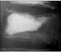 |
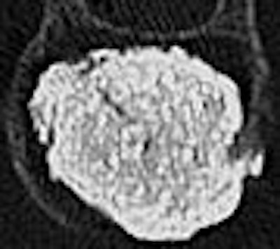
| A 73-year-old woman with vertebral compression fracture of L1 due to osteoporosis. Lateral radiograph (above) and CT scan (below) show compact and solid cement filling in vertebral body. Vertebra was assigned to type C pattern of cement distribution. Tanigawa N, Komemushi A, Kariya S, Kojima H, Shomura Y, Omura N, Sawada S, "Relationship Between Cement Distribution Pattern and New Compression Fracture After Percutaneous Vertebroplasty" (AJR 2007; 189:W1-W5). |
 |
Finally, "a new compression fracture was defined … as a vertebral body exhibiting reduced height on radiography … (or) a vertebral body exhibiting a bone marrow edema pattern on MRI," the authors explained.
The group found that a greater volume of cement was injected in patients of group C (40 vertebrae in 34 patients) than in group T (123 vertebrae in 42 patients): 4.5 ± 1.8 mL per vertebra in group C versus 3.7 ± 1.6 mL per vertebra in group T.
In group C, 27 of 34 patients had multiple fractures. This most likely occurred because a cleft indicated a cavity created by the fracture so the cement could be more easily injected, the authors stated.
In addition, those in group C had a higher incidence of new compression fractures. Also, 50% of patients with a cleft pattern had new fractures versus 26.2% of those with a trabecular pattern. An adjacent fracture was found in 44.1% of those with a cleft pattern and 16.7% of those with a trabecular pattern.
Despite these differences, VAS improvement was similar in both groups: 4.6 ± 2.5 mean pain score in group C and 4.5 ± 2.9 in group T, leading the authors to conclude that cement distribution pattern does not necessarily affect clinical response.
However, "the amount of PMMA is thought to be related to restored height of treated vertebra," the authors wrote. "We hypothesized that the restored height of a treated vertebra showing a cleft pattern is greater than that with a trabecular pattern."
This increased height may lead to an increase in soft-tissue tension around the vertebral body, which then causes an undue load on the adjacent vertebrae, they explained. Tanigawa's group also noted that the duration of fracture was higher in group C as rapid alignment restoration postprocedure may trigger new compression fractures.
By Shalmali Pal
AuntMinnie.com staff writer
December 4, 2007
Related Reading
Cortical defects on prevertebroplasty MR ups risk of cement leakage, April 3, 2007
Bone marrow edema on preop MR correlates with vertebroplasty success, May 18, 2006
Vertebroplasty increases vertebral height, imaging shows, May 2, 2006
New fractures occur sooner in adjacent vertebrae following vertebroplasty, January 24, 2006
Copyright © 2007 AuntMinnie.com


