Dear Musculoskeletal Imaging Insider,
In this issue of the Insider, we're featuring a Swiss study that found that SPECT/CT can help orthopedic surgeons and radiologists identify multiple arthritic joints in the feet and ankles and localize degenerative joint disease.
The modality also proved beneficial for surgeons and radiologists with limited clinical experience, helping them to detect the arthritic joints in small, complex anatomic sites. The study is published in the September issue of the British edition of the Journal of Bone and Joint Surgery -- learn more about its findings by clicking here.
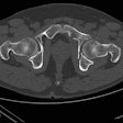
The same British journal also brings us research on MRI's ability to detect a significant number of proximal femur fractures that are missed by plain radiography, as well as prompt early diagnosis and initiation of treatment. Details from the 770-patient study come from researchers at Chelsea and Westminster Hospital in London.
Also in this issue of the Insider, read how orthopedic surgeons at the Children's Hospital of Philadelphia finally have tossed aside their hard-copy images and made the transition to digital imaging. By doing so, report turnaround time was reduced from 41 days to seven hours.
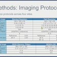
And, speaking of speed, a preliminary study has concluded that using a new 3D isotropic fast spin-echo (FSE) sequence to detect knee joint abnormalities could hasten the assessment of patients with severe pain or claustrophobia who cannot tolerate a 25-minute routine MRI exam. Researchers at the University of Wisconsin in Madison found that the 3D FSE sequence, called FSE-Cube, has similar diagnostic performance to a routine 2D MRI protocol, but takes just 20% of the time.
Keep in touch with the Musculoskeletal Imaging Digital Community in the weeks ahead, as we bring you the latest news and groundbreaking research within this specialty.


