Dear Women's Imaging Insider,
Although it is common, adenomyosis is often mistaken for other gynecological abnormalities, which can be particularly problematic when it comes to treatment. If adenomyosis is misdiagnosed as uterine fibroids, for example, uterine artery embolization (UAE) may be used as the procedure of choice. But UAE won’t solve the patient's problem because it's not targeting the right vessels, according to Dr. Edward Lyons from the University of Manitoba in Winnipeg, Canada.
In a Women’s Imaging Insider exclusive, Lyons discusses how ultrasound can help make that differential diagnosis. Lyons also explains why he believes adenomyosis is underdiagnosed, and discusses the combined role of transvaginal ultrasound and clinical and physical exams to pinpoint this disease. You can read more about it at http://www.auntminnie.com/default.asp?Sec=sup&Sub=wom&Pag=dis&ItemId=58686.
You’ll also find the latest breast imaging news in our Women’s Imaging Digital Community. This month's hot topics include the politics of mammographic screening; the usefulness of CAD for mammographic reading; and the benefits of radiotherapy to prevent cancer recurrence. Finally, we have a host of reports from the 2003 American College of Obstetricians and Gynecologists meeting. Find out how ob/gyns are incorporating imaging into their daily practice, all in our Women's Imaging Digital Community.

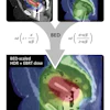


![Overview of the study design. (A) The fully automated deep learning framework was developed to estimate body composition (BC) (defined as subcutaneous adipose tissue [SAT] in liters; visceral adipose tissue [VAT] in liters; skeletal muscle [SM] in liters; SM fat fraction [SMFF] as a percentage; and intramuscular adipose tissue [IMAT] in deciliters) from MRI. The fully automated framework comprised one model (model 1) to quantify different BC measures (SAT, VAT, SM, SMFF, and IMAT) as three-dimensional (3D) measures from whole-body MRI scans. The second model (model 2) was trained to identify standardized anatomic landmarks along the craniocaudal body axis (z coordinate field), which allowed for subdividing the whole-body measures into different subregions typically examined on clinical routine MRI scans (chest, abdomen, and pelvis). (B) BC was quantified from whole-body MRI in over 66,000 individuals from two large population-based cohort studies, the UK Biobank (UKB) (36,317 individuals) and the German National Cohort (NAKO) (30,291 individuals). Bar graphs show age distribution by sex and cohort. BMI = body mass index. (C) After the performance assessment of the fully automated framework, the change in BC measures, distributions, and profiles across age decades were investigated. Age-, sex-, and height-adjusted body composition reference curves were calculated and made publicly available in a web-based z-score calculator (https://circ-ml.github.io).](https://img.auntminnie.com/mindful/smg/workspaces/default/uploads/2026/05/body-comp.XgAjTfPj1W.jpg?auto=format%2Ccompress&dpr=2&fit=crop&h=167&q=70&w=250)


