A newly updated chest x-ray imaging score could be a valuable tool for measuring the severity of COVID-19 pneumonia, according to research published online December 5 in Scientific Reports.
A group at the University of Copenhagen in Denmark tweaked a tool first developed by Italian clinical researchers in 2020 called the Brixia score. In a test, they found modified Brixia (MBrixia) scores were associated with higher needs of respiratory support in COVID-19 patients.
"The MBrixia score could be implemented as a supplementary tool in routine clinical care of COVID-19 patients," wrote first author Christian Jensen and colleagues.
CT has a higher sensitivity for detecting COVID-19 than chest x-ray, yet chest x-ray is comparatively inexpensive and readily available. Moreover, portable chest x-ray can be used without the need for in-hospital transportation of potentially infectious patients, according to the researchers.
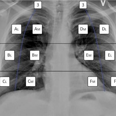
The Brixia score represents the sum of a rule-based quantification of chest x-ray on a scale of zero to three in 12 anatomical zones in the lungs. Italian researchers first developed the tool in 2020 in a study showing it could be useful for predicting in-hospital mortality in patients with COVID-19.
In this study, the definition of a score of three was altered from indicating interstitial and alveolar infiltrates with alveolar predominance to consolidations, which may more accurately reflect the trajectory of COVID-19 pneumonia, the researchers wrote.
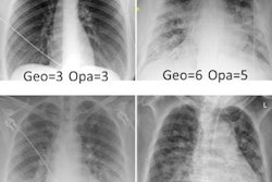
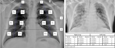 (A) The MBrixia scoring system. Box 1 and 2 mark the horizontal division corresponding to the inferior wall of the aortic wall, and the inferior wall of the right pulmonary vein, respectively, creating three zones in each lung: A, B, C, D, E and F. Box 3 marks the vertical line drawn from the pulmonary apices to the diaphragm, creating further division into medial (M) and lateral (L) zones. (B) A clinical example of a chest x-ray image scored using the MBrixia score. Image courtesy of Scientific Reports through CC BY 4.0 International License.
The group applied the MBrixia score to 290 chest x-ray images acquired from 37 COVID-19 patients between May 4 and June 5, 2020. Twenty-two (59.5%) patients were admitted to the intensive care unit and 10 (27%) died during follow-up. Respiratory support was categorized according to severity based on an ordinal scale of five levels.
According to the analysis, higher MBrixia scores were associated with higher levels of respiratory support at the time chest x-rays were performed. Specifically, when compared with patients receiving no supplemental oxygen, median MBrixia scores of COVID-19 patients were between 1.75 and 2.38 times higher for patients on lower levels of oxygen support and between 2.66 and 3.52 times higher for patients requiring invasive mechanical ventilation or extracorporeal membrane oxygenation (ECMO).
"The MBrixia score's modified definition... made it possible to capture a more detailed spatial quantification of COVID-19 pneumonia," the authors wrote.
Ultimately, with future validation, the use of MBrixia score in the clinical setting could prove beneficial in tracking progression of COVID-19 pneumonia over time, the group suggested.
"Future studies should investigate the score's validity and capabilities of predicting clinical outcomes," the group concluded.

